| ON THE COVER | 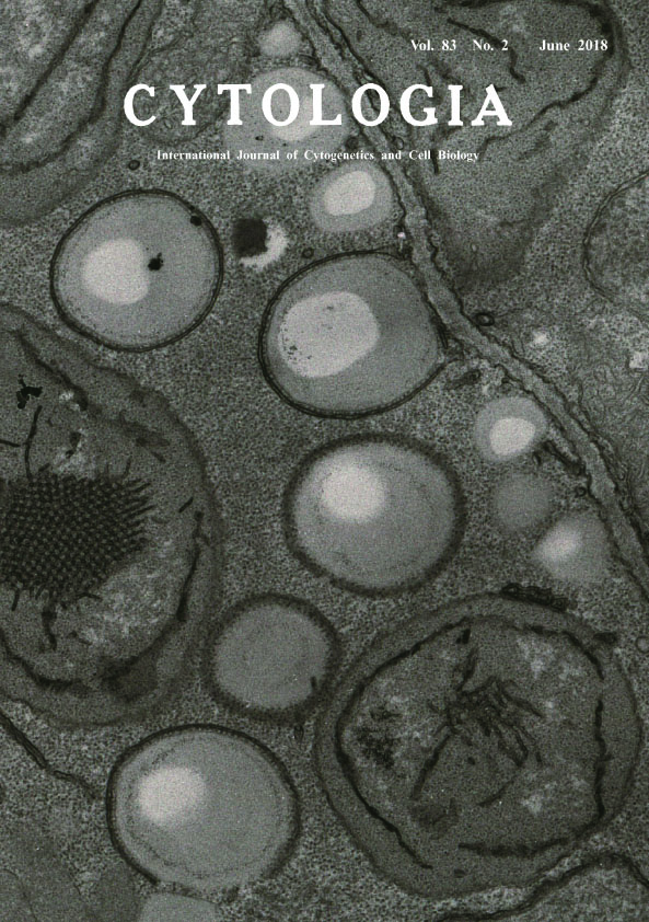 |
|
|---|---|---|
| Vol. 83 No.2 June 2018 | ||
| Technical Note | ||
|
|
||
Existence of Lipid Bodies Surrounded by Membranes in Early Greening Cotyledonary Cells Yasuko Hayashi1*, Chinatsu Takagi1 and Mikio Nishimura2 1 Department of Environmental Science, Graduate School of Science and Technology, Niigata University, Niigata 950–2181, Japan 2 Department of Cell Biology, National Institute for basic Biology, Okazaki, Aichi 444–8585, Japan Received November 8, 2017; accepted December 10, 2017 Lipid bodies (LBs) are cytosolic lipid droplets described in plants and all other eukaryotes. They are dynamic organelles controlling neutral lipid metabolism and storage. In oil seed plants, LBs are essential for reproduction because they store oil in seeds, which are an important source of energy during post-germinative growth until the acquisition of photo-autotrophism. We and other scientists have investigated organelle interactions during post- germinative growth under the electron microscope, and sometimes recognized the existence of membranes near LBs; however, these membranes have not yet been investigated. In this research, we attempted chemical fixation with osmium tetroxide and potassium ferrocyanide to make the observation of membranes under the electron microscope possible. Samples were Arabidopsis thaliana cotyledons grown in the dark for 5 days after germination and irradiated with light for 30 min. Samples were fixed for 3 h at 4°C in cacodylate buffer (pH 7.4) containing 4% paraformaldehyde, 1% glutaraldehyde, and 0.1 M CaCl2. Samples were washed seven times with 0.1 M cacodylate buffer for 1.5 h and postfixed in 0.1 M cacodylate buffer containing 2% OsO4, 0.8% K3Fe(CN)6, and 1 μM CaCl2 for 2 h at room temperature. Samples were washed seven times with distilled water for 1.5 h and dehydrated with ethanol series, then embedded in Spurr resin. Ultrathin sections were cut with a diamond knife on an ultramicrotome and mounted on a Formvar-coated grid. Samples were stained with uranium acetate and lead and then observed using an electron microscope (H-7650, Hitachi) at 80 kV. In the cotyledonary cells, we recognized many naked LBs and LBs associated with membranes, and revealed that some LBs are completely surrounded by membranes rather than having partial contact. One possibility for the identity of the membrane is the endoplasmic reticulum (ER). The relationship between LBs and the ER has been reported not only in plants, animals, and yeast but also in green algae (Kuroiwa et al. 2014). As it is known that the ER is involved in the formation of LBs and the accumulation of lipids, the presence of a relationship between LBs and the ER in the cotyledon of developing seeds is reasonable. Recently, the involvement of the ER in lipid storage in aging leaves was reported (Brocard et al. 2017). However, this relationship between LBs and the ER in postgerminative cotyledon growth is strange because during this period, storage lipids could be consumed for product energy. This phenomenon may indicate a newly identified function of the ER in cotyledonary cells. Another possibility is that it is an autophagic isolation membrane. As surplus storage lipids become unnecessary in the greening of cells that are ready for photosynthesis, cells may start the process of lipophagy.
Brocard, L., Immel, F., Coulon, D., Esnay, N., Tuphile, K., Pascal, S., Claverol, S., Fouillen, L., Bessoule, J. and Brehelin, C. 2017. Proteomic analysis of lipid droplets from Arabidopsis aging leaves brings new insight into their biogenesis and functions. Front. Plant Sci. 8: 894. Kuroiwa, T., Ohnuma, M., Imoto, Y. and Kuroiwa, H. 2014. Lipid droplet formation in cells of the filamentous green alga Klebsormidium nitens as revealed by BODIOY-DiOC6 and BODIPY-Nile red double-staining microscopy. Cytologia 79: 501–507. *Corresponding author, e-mail:yhayashi@env.sc.niigata-u.ac.jp DOI: 10.1508/cytologia.83.123 |
||