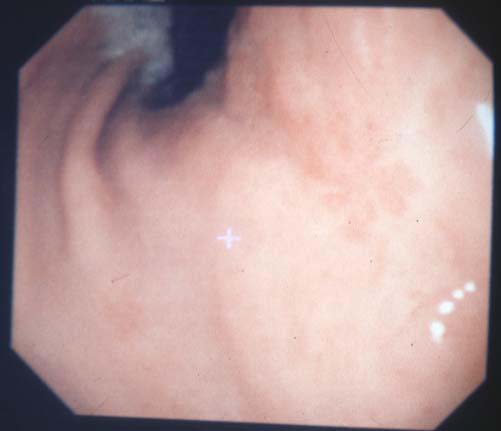
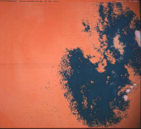
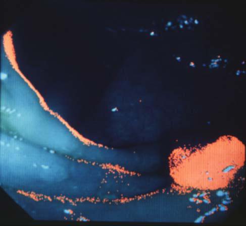
パネルディスカッション 第2部:医療の最前線におけるデジタル画像の活用とその色処理 消化器内視鏡診療の立場から 勝 健一 大阪医科大学第2内科 The 1st Symposium of the 'Color' of Digital Imaging in Medicine Panel Discussion Part 2 : Digital Imaging and Its Color Management in the Medical Forefront From the Standpoint of Gastrointestinal Endoscopy Ken-ichi KATSU Department of 2nd Department of Internal Medicine, Osaka Medical College Summary Introduction of digital imaging with electronic endoscopy is expected to realize (1) computerized endoscopic diagnosis based on digitized image data, (2) computerized laser cauterization on the basis of automatic detection and targeting of early cancer and (3) real time superimposition of various referential data on endoscopic images. Using electronic endoscopes and an image analysis system, digitized color data were collected from gastric mucosa of normal control, atrophic gastritis and early cancer and analyzed in respect of the difference in color. Statistical analysis could not discern any significant difference in color among these mucosa, but primary wavelength of light which comes from mucosa showed a significant difference between the normal one and the others. Besides, the affected area of mucosa could be detected and made visible by computerized image processing in one case of early cancer of the IIc type (Fig. 1, Fig. 2), and in one case of early colon cancer of an elevated type (Fig. 3), though in most cases this was not possible. Further investigation and improvement of instruments are indicated to pursue the possibilities of digital imaging in gastrointestinal endoscopy, including the application of telemedicine and development of a referential case database for endoscopic diagnosis. |


4.考 察 現在のところ内視鏡画像のコンピュータ自動画像診断に関する成果は以上しか成功していないが,恐らく世界で初めての画像と分析データと確信している。 内視鏡画像として明らかに判別できる病変がコンピュータによる波長解析で明確な診断的優位性を示さなかったのはヒトの色弁別能力を超えた解析ソフトでなかったためと考えられる。すなわち,Wrightらによればヒトは 490~590nm 近傍では1nmの波長弁別能力があると報告している。Bedford は視野角の狭小により判別力の低下を報告しているがそれでも論文中の図から推定すると4nm 程度しか低下していない。この事はヒトの色判別力の精度の高さに比較して現在まで使用していたパーソナルコンピュータと測定器機の精度が不十分であったためと考えられる。しかし,1症例であったが色差の抽出により早期胃癌の病巣部分を描出できたことはコンピュータ診断における病巣描出の可能性が期待される。今後,多くの症例において恒常的に病巣部を描出するには,そのための条件を解明しなければならない。電子内視鏡によるコンピュータ診断の理想としては癌などの病変に特有な波長を発見し,これをマーカーとして検索することである。また,同一画像内において推計学的に有意差をしめす主波長の部分を検索すれば早期胃癌か萎縮性胃炎,さらに癌病巣を画像として抽出できることが示唆された。さらに大腸内視鏡で発見された隆起型早期癌に於いて色差抽出法で同部が擬似カラーで表示されて(図3)おり,今後,症例の蓄積と機器のバージョンアップによりデジタル化電子内視鏡画像によるコンピュータ診断が可能となると考えている。 図3
|

また,電子内視鏡像の無線伝送については前任地埼玉医科大学では内視鏡検査室において
UHF
のテレビ周波を用いて電子内視鏡の映像出力を発信し,通常のテレビおよび携帯テレビで検査中の内視鏡映像を学生達に供覧していた。将来はデジタル通信,通信衛星などにより地球上のいかなる地域で内視鏡検査をうけてもリアルタイムであるいは保存画像を,たとえば国立がん診断支援センターのような解析センターからのセカンドオピニオンや内視鏡診断書が得られる時代がくることを想定しての実験であった。当面はLAN,ISDNなどの全国的通信網によりデータファイルの蓄積を行ない,普遍的な診断を可能とすることが必要である。さらにデジタル診断とリアルタイム対応により検査時に内視鏡直視下に病巣を内視鏡レーザー焼灼システムとコンピュータと連動させることを期待している。すなはち,スクリーニング検査時に微小早期がんを自動的に焼灼することにより消化管進行癌の予防を期待している。また,画像内に心電図,内視鏡直視下の粘膜表面pHおよび粘膜電位(ポテンシアルデイファレンス),血流測定, 粘膜の硬さ(g/cm),温度,粘膜断面超音波像(Bモード)などの生理学的測定値を表示させ,胃潰瘍・粘膜下腫瘍や潰瘍性大腸炎の早期発見の可能性も期待している。
|