| ON THE COVER | 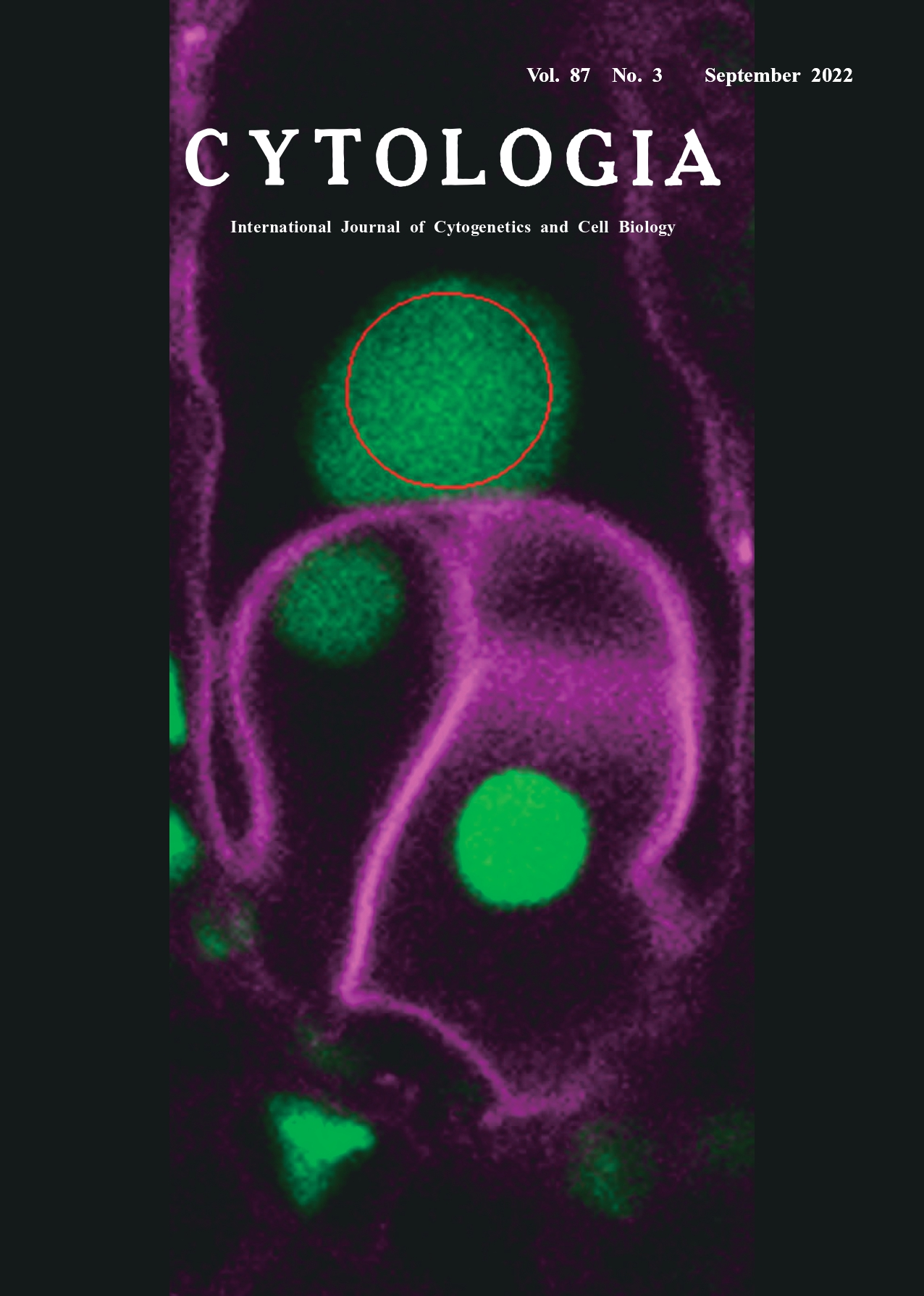 |
||
|---|---|---|---|
| Vol. 87 No.3 September 2022 | |||
| Technical Note | |||
|
|
|||
Deep Imaging and Optical Manipulation of Double Fertilization Shiori Nagahara1*, Tetsuya Higashiyama2* 1 Institute of Transformative Bio-Molecules, Nagoya University, Furo-cho, Chikusa-ku, Nagoya, Aichi 464–8601, Japan 2 Department of Biological Sciences, Graduate School of Science, The University of Tokyo, 7–3–1 Hongo, Bunkyo-ku,Tokyo 113–0033, Japan
Technical innovations to microscopy have allowed functional imaging of various biological phenomena (Higashiyama et al. 2021). In addition to visualization improvements, laser-based technologies permit the optical manipulation of specific cells in living tissues and organs. Here, we show optical manipulation of double fertilization in Arabidopsis thaliana, which occurs deep inside the flower organs Angiosperm reproduction involves the growth of male pollen tubes into the pistil where two male gametes are transported to the ovules. In the ovule, the two sperm cells are discharged from the pollen tube to fertilize distinct female gametes (an egg cell and a central cell). The fertilized egg and central cell give rise to the embryo and endosperm, respectively. This process is called double fertilization. The inaccessibility of the female gametophyte embedded in the pistil makes direct observation of this event challenging. Two-photon excitation microscopy (2PEM) equipped with a multiphoton laser constitutes a key approach for visualization and analysis of structures deep within flowering plants (Mizuta 2021). The near-infrared femtosecond pulse laser (multiphoton laser) of 2PEM is less invasive and can deeply penetrate biological tissues. Thus, 2PEM allows for deep-tissue imaging in living ovules, including subcellular visualization of the dividing zygote of an Arabidopsis ovule (e.g., Kimata et al. 2016). In addition, highly focused multiphoton lasers can disrupt cells embedded in the ovule. Multiphoton laser irradiation of the apical cell of a 1-cell embryo successfully disrupted the apical cell without lethal damage to the remaining basal cell and surrounding female tissue (Gooh et al. 2015). The cover image shows multiphoton laser irradiation of a central cell nucleus in the ovule of A. thaliana. We specifically damaged one of the two fertilization targets of sperm cells (egg or central cells) without disrupting other ovular cells, to address whether defects in one target increased polyspermy in the other target. The ovule was excised from the pistil of a transgenic plant possessing RPS5Ap::H2B-GFP and RPS5Ap::td-Tomato-LTI6b genes, which label nuclei and plasma membrane, respectively. The ovule was incubated with pollen tubes on pollen germination media and laser irradiation of an individual female gamete was performed after 2–3 h of incubation. An 880 nm multiphoton laser (Chameleon Vision II, Coherent, Santa Clara, CA, USA) equipped with a confocal microscope (LSM780-DUO-NLO, Zeiss, Oberkochen, Germany) was used to irradiate the central cell nucleus (red circle). Then, the laser-irradiated ovule that would normally receive released sperm was analyzed (Nagahara et al. 2021). Single fertilization of the central cell was significantly increased in the egg cell irradiated ovules, implying that disruption of the egg cell was successful in most cases and that the mechanism to accept only one sperm cell (i.e., polyspermy block) is working in the central cell, although it was less strict than that in the egg cell. Deep imaging and optical manipulation using 2PEM can accelerate our understanding of cell-to-cell communication in tissues involved in angiosperm reproduction.
Gooh, K., Ueda, M., Aruga, K., Park, J., Arata, H., Higashiyama, T. and Kurihara, D. 2015. Live-cell imaging and optical manipulation of Arabidopsis early embryogenesis. Dev. Cell 34: 242–251. Higashiyama, T., Maizel, A. and Simon, R. 2021. Seeing is believing: Advances in plant imaging technologies. Plant Cell Physiol. 62: 1217–1220. Kimata, Y., Higaki, T., Kawashima, T., Kurihara, D., Sato, Y., Yamada, T., Hasezawa, S., Berger, F., Higashiyama, T. and Ueda, M. 2016. Cytoskeleton dynamics control the first asymmetric cell division in Arabidopsis zygote. Proc. Natl. Acad. Sci. U.S.A. 113: 14157–14162 Mizuta, Y. 2021. Advances in two-photon imaging in plants. Plant Cell Physiol. 62: 1224–1230. Nagahara, S., Takeuchi, H. and Higashiyama, T. 2021. Polyspermy block in the central cell during double fertilization of Arabidopsis thaliana. Front. Plant Sci. 11: 588700. * Corresponding author, e-mail: nagahara.shiori.j0@f.mail.nagoya-u.ac.jp; higashi@bs.s.u-tokyo.ac.jp DOI: 10.1508/cytologia.87.201 |
|||