| ON THE COVER | 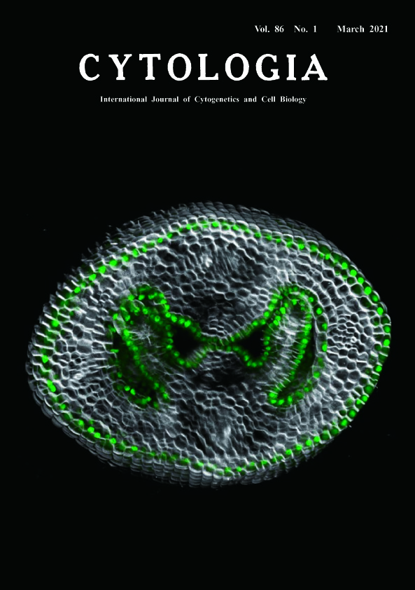 |
||
|---|---|---|---|
| Vol. 86 No.1 March 2021 | |||
| Technical Note | |||
|
|
|||
Postgenital Fusion and Epidermal Cell Fate Control during Gynoecium Development Aika Yokoi1 and Mitsuhiro Aida2* 1 Graduate School of Biological Sciences, Nara Institute of Science and Technology, 8916–5 Takayama, Ikoma, Nara 630–0192, Japan 2 International Research Organization for Advanced Science and Technology, Kumamoto University, 2–39–1 Kurokami, Chuo-ku, Kumamoto 860–8555, Japan
Flowering plants develop a hollow and closed reproductive structure called the gynoecium (Ferrandiz et al. 2010). Formation of the gynoecium often involves a characteristic developmental process called postgenital fusion, in which protodermal cells of two or more discrete primordia adhere with one another along their surface cell walls. In Arabidopsis thaliana, for example, a pair of septum primordia undergo postgenital fusion to form a single septum that separates the gynoecium into two locules (Roeder and Yanofsky 2006). To gain insight into the mechanism of cell fate control during septum fusion, we examined the expression of ARABIDOPSIS THALIANA MERISTEM LAYER 1 (ATML1), a regulatory gene that promotes epidermal cell fate in shoot organs (Abe et al. 2003), using the pATML1::NLS:3xEGFP reporter (Takada and Jürgens 2007). Gynoecia were excised from stage 10 flowers and embedded in 1% agar blocks, which were subsequently subjected to fixation and clearing as per the ClearSee method (Kurihara et al. 2015). Sample blocks were stained with 100 μg mL-1 Calcofluor White M2R (Sigma-Aldrich, STL, USA) to visualize the cell walls and subjected to imaging using a Leica TCS SPE confocal microscope (Leica Microsystems, Wetzlar, Germany). A stack of 45 optical transverse sections was acquired with a 0.5-μm step size, and a three-dimensional (3D) volume image was reconstructed using the Leica LAS X software (Leica Microsystems). In the reconstructed 3D image (cover figure), a pair of septum primordia was observed as two opposing domeshaped structures that touch one another at their distalmost portions, and the GFP signal (green) was detected in the protodermal layer of the two primordia. Importantly, cells at their contacting surfaces also accumulated GFP, indicating that these cells retained an epidermal character. Our results indicate that the septum cells that undergo postgenital fusion possess an epidermal character that is marked by ATML1 expression even after they have touched one another. Further characterization of ATML1 expression and function during septum formation would be important to understand the mechanisms of postgenital fusion and epidermal cell fate control. We thank Shinobu Takada for providing seeds of the ATML1 reporter, José Irepan Reyes-Olalde for critically reading the manuscript, and Enago (www.enago.jp) for the English language review.
Abe, M., Katsumata, H., Komeda, Y. and Takahashi, T. 2003. Regulation of shoot epidermal cell differentiation by a pair of homeodomain proteins in Arabidopsis. Development 130: 635–643. Ferrandiz, C., Fourquin, C., Prunet, N., Scutt, C. P., Sundberg, E., Trehin, C. and Vialette-Guiraud, A. C. M. 2010. Carpel Development. In: Kader, J. C. and Delseny, M. (eds.). Advances in Botanical Research Vol. 55. Academic Press Ltd-Elsevier Science Ltd., London. pp. 1–73. Kurihara, D., Mizuta, Y., Sato, Y. and Higashiyama, T. 2015. Clear- See: A rapid optical clearing reagent for whole-plant fluorescence imaging. Development 142: 4168–4179. Roeder, A. H. and Yanofsky, M. F. 2006. Fruit development in Arabidopsis. Arabidopsis Book 4: e0075. Takada, S. and Jürgens, G. 2007. Transcriptional regulation of epidermal cell fate in the Arabidopsis embryo. Development 134: 1141–1150. * Corresponding author, e-mail: m-aida@kumamoto-u.ac.jp DOI: 10.1508/cytologia.86.1 |
|||