| ON THE COVER | 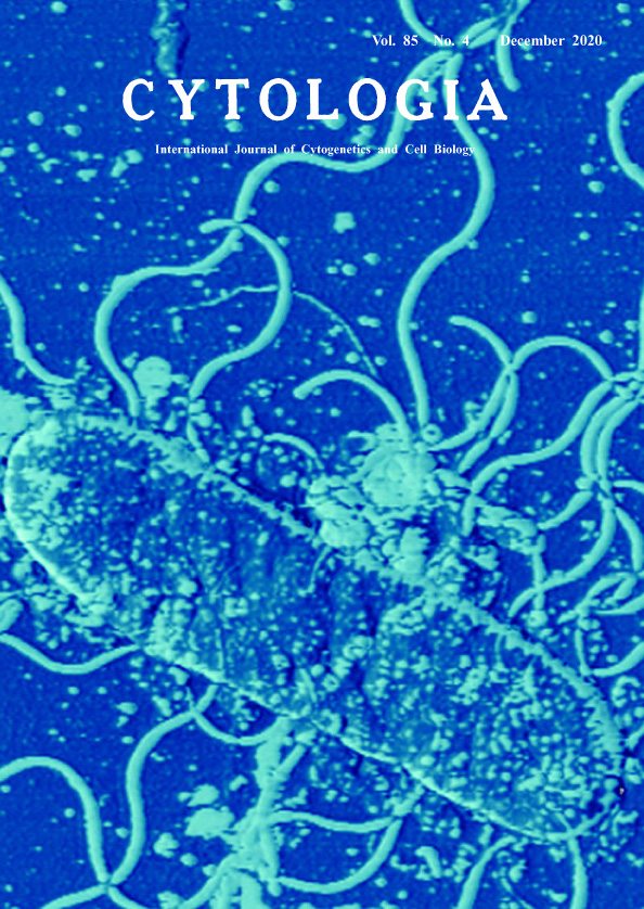 |
||
|---|---|---|---|
| Vol. 85 No.4 December 2020 | |||
| Technical Note | |||
|
|
|||
Visualization of Bacteria (Vibrio alginolyticus) as They Really Are by Atomic Force Microscopy Susumu Yoshizawa1,2*, Eiko Ikemoto1 and Kazuhiro Kogure1 1 Atmosphere and Ocean Research Institute, The University of Tokyo, Chiba 277–8564, Japan 2 Graduate School of Frontier Sciences, The University of Tokyo, Chiba 277–8563, Japan
Bacteria are tiny and cannot be observed by the human eye, therefore using a microscope is essential for observing their morphology. However, bacteria are too small to visualize their actual shape using conventional fluorescence microscopy. Therefore, we use atomic force microscopy (AFM) to observe the exact shape of bacteria. Besides, because the polar and lateral flagella of bacteria are indicators of their characteristics, visualization of their actual shape can help us to understand their ecology. The cover image showed the bacteria Vibrio alginolyticus YM19) observed by AFM (model SPM-9500 J2; Shimadzu). V. alginolyticus YM19 was the flagellumdefective mutant (polar flagella- and lateral falagella+) (Atsumi et al. 1996). The mutant bacteria were grown in 1/2 strength ZoBell 2216E medium prepared with 80% aged seawater at 25°C using a rotary shaker. Bacterial cells in their late exponential growth phase were fixed with formaldehyde (final conc. 2%). Cells were concentrated on a filter (Isopore filter, pore size 0.2 μm, Whatman). The observations were obtained with atomic force microscopy (SPM-9500 J2, Shimadzu Col Ltd.) in dynamic mode. The instrument was equipped with a microfabricated and oxide-sharpened Si3N4 cantilever (OMCL-AC160TS-C1, Olympus Co. Ltd.) that has a pyramidal tip and has force with a spring constant of 42 N m-1. The detailed methods were described previously (Nishino et al. 2004). Our group has used the AFM to observe a variety of bacteria. For example, we have used images taken with AFM to characterize bacterial shape in research papers proposing new species of luminous bacteria (Yoshizawa et al. 2009, 2012). It is because we need to know their exact shape of bacteria to understand their real ecology. The images taken so far are shown on the following website. If you are interested in these images, please visit this site. “Observing microorganisms as they are.” Website URL: https://sites.google.com/view/aori-microbe-afm/home
Atsumi, T., Maekawa, Y., Yamada, T., Kawagishi, I., Imae, Y. and Homma, M. 1996. Effect of viscosity on swimming by the lateral and polar flagella of Vibrio alginolyticus. J. Bacteriol. 178: 5024–5026. Nishino, T., Ikemoto, E. and Kogure, K. 2004. Application of atomic force microscopy to observation of marine bacteria. J. Oceanogr. 60: 219–225. Yoshizawa, S., Tsuruya, Y., Fukui, Y., Sawabe, T., Yokota, A., Kogure, K., Higgins, M., Carson, J. and Thompson, F. L. 2012. Vibrio jasicida sp. nov., a member of the Harveyi clade, from marine animals (packhorse lobster, abalone, and Atlan Yoshizawa, S., Wada, M., Kita-Tsukamoto, K., Ikemoto, E., Yokota, A. and Kogure, K. 2009. Vibrio azureus sp. nov., a luminous marine bacterium isolated from seawater. Int. J. Syst. Evol. Microbiol. 59: 1645–1649. * Corresponding author, e-mail: yoshizawa@aori.u-tokyo.ac.jp DOI: 10.1508/cytologia.85.261 |
|||