| ON THE COVER | 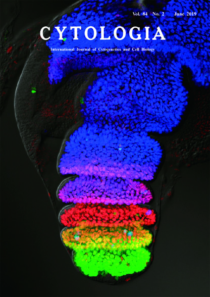 |
|
|---|---|---|
| Vol. 84 No.2 June 2019 | ||
| Technical Note | ||
|
|
||
Subdivision of the Tarsal Region into Five Tarsal Segments by the Combination of Region-Specific Transcription Factors Kohei Natori and Tetsuya Kojima* Graduate School of Frontier Sciences, The University of Tokyo, 5–1–5 Kashiwanoha, Kashiwa, Chiba 277–8562, Japan Received November 5, 2018; accepted January 29, 2019 The adult leg of the fruit fly, Drosophila melanogaster, consists of several segments along the proximodistal axis, which are, from proximal to distal direction, the coxa, trochanter, femur, tibia, tarsus and pretarsus. The tarsus is further subdivided into five subsegments and they are called the tarsal segment 1 (ta1), the tarsal segment 2 (ta2), the tarsal segment 3 (ta3), the tarsal segment 4 (ta4) and the tarsal segment 5 (ta5) with ta1 being the most proximal segment. The mechanism of subdividing the tarsal region into five regions corresponding to each tarsal segment has been extensively studied (Kojima 2017). In addition, the number of tarsal segments is highly variable among insect species, so that the tarsal development is a good model for studying morphological evolution and diversification. To see the subdivision of the tarsal region during leg development in D. melanogaster, the leg primordia was dissected from the prepupa of the Canton-S strain at 4 h after puparium formation and simultaneously immunostained with antibodies against region-specific transcription factors as described previously (Natori et al. 2012). The rabbit anti-BarH1/BarH2 (Bar) antibody (Higashijima et al. 1992), rat anti-Bric à brack (Bab1) antibody (Couderc et al. 2002) and mouse anti-Dachshund (Dac) antibody (Mardon et al. 1994) were used as first antibodies. Alexa 488 conjugated anti-rabbit IgG antibody (Molecular Probe), Cy3 conjugated anti-rat IgG antibody (Jackson Immune Research) and Alexa 647 conjugated anti-mouse IgG antibody (Molecular Probe) were used as secondary antibodies. Images were acquired using the confocal microscope FV1000 (Olympus). The expression of Bar, Bab1 and Dac are shown in green, red and blue, respectively, in the image of cover photograph. Strong and weak Bar expression (green) are observed in ta5 and ta4, respectively, while Dac is expressed weakly in ta2 and strongly in ta1 and more proximal segments. In the intermediate region, ta2–ta4 evidently express Bab1. The image demonstrates the beautiful “color-coding” in the tarsal region and shows that each tarsal segment is distinguished from other tarsal segments by the specific combination of the region-specific transcription factors: Bar only (ta5; green), Bar and Bab1 (ta4; yellow), Bab1 only (ta3; red), Bab1 and Dac (ta2; magenta) and Dac only (ta1; blue).
Couderc, J. L., Godt, D., Zollman, S., Chen, J., Li, M., Tiong, S., Cramton S. E., Sahut-Barnola, I. and Laski, F. A. 2002. The bric à brac locus consists of two paralogous genes encoding BTB/ POZ domain proteins and acts as a homeotic and morphogenetic regulator of imaginal development in Drosophila. Development 129: 2419–2433. Higashijima, S., Kojima, T., Michiue, T., Ishimaru, S., Emori, Y. and Saigo, K. 1992. Dual Bar homeo box genes of Drosophila required in two photoreceptor cells, R1 and R6, and primary pigment cells for normal eye development. Genes Dev. 6: 50–60. Kojima, T. 2017. Developmental mechanism of the tarsus in insect legs. Curr. Opin. Insect Sci. 19: 36–42. Mardon, G., Solomon, N. M. and Rubin, G. M. 1994. dachshund encodes a nuclear protein required for normal eye and leg development in Drosophila. Development 120: 3473–3486. Natori, K., Tajiri, R., Furukawa, S. and Kojima, T. 2012. Progressive tarsal patterning in the Drosophila by temporally dynamic regulation of transcription factor genes. Dev. Biol. 361: 450–462. *Corresponding author, e-mail:tkojima@k.u-tokyo.ac.jp DOI: 10.1508/cytologia.84.105 |
||