| ON THE COVER | 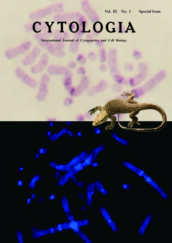 |
|
|---|---|---|
| Vol. 82 No.1 Special issue 2017 | ||
| Technical Note | ||
|
|
||
Constitutive Heterochromatin Observed on Metaphase Chromosome of Varanus bengalensis by C-Banding and DAPI Methods Isara Patawang1 and Alongklod Tanomtong2* 1Department of Biology, Faculty of Science, Chiang Mai University, Chiang Mai, Muang 50200, Thailand 2Toxic Substances in Livestock and Aquatic Animals Research Group, Department of Biology, Faculty of Science,Khon Kaen University, Khon Kaen, Muang 40002, Thailand Received October 12, 2016; accepted December 30, 2016 Key words Constitutive heterochromatin, Varanus bengalensis, C-banding technique, DAPI method. Constitutive heterochromatin, mainly formed at the gene-poor regions of pericentromeres, is a condensed DNA region found throughout the vertebrate chromosomes and other eukaryotes. Pericentromeric regions comprise of repetitive tandem sequences that are precious in chromosome segregation of mitotic division. For the C-banding technique of the present experiment, we treat the chromosomes with 0.2 N HCl for 15 min at room temperature and 0.05 N Ba(OH)2 for 15 min at 37°C to break the histone protein throughout the euchromatin and loosen the heterochromatin, and stain them with 20% Giemsa solution for 30 min. Our result showed the appearance of constitutive heterochromatin in various regions on each chromosome that included centromeric, subcentromeric, some interstitial and some telomeric regions (Patawang et al. 2017). The DAPI staining was made by the standard method of FISH technique; chromosomes were digested by 10 mM HCl and pepsin, from porcine gastric mucosa, for five minutes at 37°C and then stabilized with a postfixsolution (2% paraformaldehyde, 1× PBS and 1 M MgCl2 solution) for 10 min at room temperature and stained with antifade-4′,6-diamidino-2-phenylindole solution in a dark chamber. The DAPI band chromosome was captured by fluorescence microscope CFI 60 infinity optical lighting system. The picture on the title page shows the constitutive heterochromatin regions by the two techniques that reveal homology band from both C-banding and DAPI methods. The large clearly condensed heterochromatins appear on the first and second pairs of macrochromosomes. In addition, the regions of subcentromeres of many microchromosome were alike, found to exhibit the same pattern by the two methods. The chromosomal character by the C-banding method has the dark purple band of Giemsa dye that stained the histone protein of constitutive heterochromatin, while the DAPI band showed the bright blue on various repetitive tandem repeat areas of each chromosome. An antifade-DAPI binds to a minor groove of DNA, especially an A–T rich region that is often found in repetitive tandem repeats of constitutive heterochromatin. The C-banding and DAPI methods have the same final result of constitutive heterochromatin appearing, but differ in the staining mechanism of the dyes.
Patawang, I., Tanomtong, A., Getlekha, N., Phimphan, S., Pinthong, K. and Neeratanaphan, L. 2017. Standardized karyotype and idiogram of Bengal monitor lizard, Varanus bengalensis (Squamata, Varanidae). Cytologia 82: 75–82. *Corresponding author, e-mail: tanomtong@hotmail.com DOI: 10.1508/cytologia.82.1 |
||