| ON THE COVER | 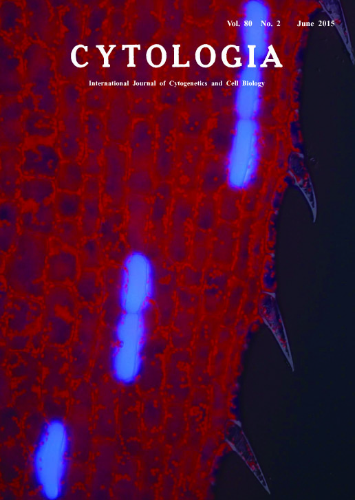 |
|
|---|---|---|
| Vol. 80 No.2 June 2015 | ||
| Technical note | ||
|
|
||
| Observation of Colorless Idioblasts in Egeria densa Leaves by Conventional Ultraviolet-Excitation Fluorescence Microscopy Makoto T. Fujiwara1,*, Emi Kobayashi1, Mikako Kanazawa1 and Ryuuichi D. Itoh2 Over the history of botany, the exploration of plant materials with remarkable morphologies or phenomena was crucial for scientific education, as well as research. The development of laboratory experiments for middle education or introductory courses of higher education often requires conditions to be met with regard to the cost of instruments, the availability of organisms, and the clarity of experiment outcomes, which are distinct from those of frontier research. Egeria densa Planch., a submerged freshwater macrophyte, satisfies the above criteria in many aspects, allowing its wide use in cytological and physiological experiments such as observations of photosynthetic oxygen evolution, cell structure, and plasmolysis/deplasmolysis. Following its introduction from the United States in the 1920s with an erroneous designation of ‘Elodea canadensis’, this species has become a popular aquarium plant in Japan. One of the first studies in Japan to use this organism was reported by Kogane Kiyohara in Cytologia (Kiyohara 1930). In a recent study, we revealed the differentiation pattern of chloroplast-deficient idioblast cells of E. densa leaves (Hara et al. 2015). Egeria idioblasts are visually transparent among the surrounding chloroplast-abundant epidermal cells but have drawn little attention since their description by Hans Solereder a century ago (1913; cited in our paper). Upon irradiation of mature leaves with ultraviolet light, idioblasts emitted bright light-blue fluorescence, while normal epidermal cells emitted red fluorescence from chloroplasts (see Cover Image). Excitation of unknown molecules that accumulate in vacuoles might cause fluorescence from the entire area of idioblasts. This observation was a rediscovery of the original work by Ernst Perner (Perner 1950) and achieved using a conventional U-excitation filter set and epifluorescence microscopy even at a low magnification, revealing the unique distribution of idioblasts at the whole-leaf level. Microscopic sights obtained by a simple methodology might inspire future, as well as current, scientists to sense the issues of cell differentiation, patterning, and metabolism, expanding the utility of this plant in laboratory experiments.
Perner, E. S. 1950. Die intravitale Fluorochromierung junger Blätter von Helodea densa. Protoplasma 39(3): 400-422. Hara, T., Kobayashi, E., Ohtsubo, K., Kumada, S., Kanazawa, M., Abe, T., Itoh, R. D. and Fujiwara, M. T. 2015. Organlevel analysis of idioblast patterning in Egeria densa Planch. leaves. PLoS ONE 10(3): e0118965. 1 Department of Biology, Sophia University, 7-1 Kioicho, Chiyoda-ku, Tokyo 102-8554, Japan, 2Department of Chemistry, Biology and Marine Science, Faculty of Science, University of the Ryukyus, Senbaru 1, Nishihara, Okinawa 903-0213, Japan. *Corresponding author, e-mail: mtf1@mac.com DOI: 10.1508/cytologia.80.131 |
||