| ON THE COVER | 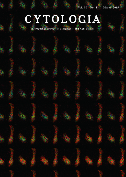 |
|
|---|---|---|
| Vol. 80 No.1 March 2015 | ||
| Technical note | ||
|
|
||
| Real Time Imaging of Biological Phenomena with Super-duper Luminescent Proteins Kenta Saito1, Yu-Fen Chang1, Kazuki Horikawa2, Yuriko Higuchi3, Mitsuru Hashida3,Tomoki Matsuda1, Yoshiyuki Arai1, Takeharu Nagai1 and Takeharu Nagai1* Fluorescent protein technology revolutionized our understanding of biological processes. However the requirement for external illumination definitely precludes its universal application to all biological processes in all tissues. On the other hand, bioluminescent proteins such as luciferase don’t require the external illumination, by which bioluminescence imaging can be acquired without phototoxicity and autofluorescence from the specimen, allowing us signal detection with high signal-to-noise ratio. These properties make bioluminescent proteins potentially superior to fluorescent proteins as a bioimaging tool. However, bioluminescence signals are too dim to be measured in real time, requiring longer exposure than fluorescence imaging that takes less than 1 s. To overcome this drawback, Renilla reniformis luciferase (Rluc) was conducted on random mutagenesis to improve the intensity. Then, the luminescence intensity was further increased by fusion of the improved Rluc to Venus, a yellow fluorescent protein variant, with high FRET efficiency. The chimeric protein named Nano-lantern showed much brighter luminescence than the commercially available Rluc, enabling not only real-time imaging of intracellular structures in living cells with spatial resolution equivalent to fluorescence but also sensitive tumor detection in freely moving mice which has never been possible before (Saito et al. 2012) . We then made a Ca2+ indicator named Nano-lantern (Ca2+) by insertion of a Ca2+- sensing domain (calmodulin-M13) into the Rluc moiety of Nano-lantern. Nano-lantern (Ca2+) showed a 300% signal change upon Ca2+ binding, and enough bright to perform video-rate (30 frame/s) imaging, thereby we succeeded imaging Ca2+ entry upon ChR2 activation with blue light (Arai and Nagai 2014). These super-duper luminescent proteins will revolutionize conventional bioimaging by allowing visualization of biological phenomena not seen before at the single-cell, organ, and whole-body level, in animals and plants.
Saito, K., Chang, Y. F., Horikawa, K., Hatsugai, N., Higuchi, Y., Hashida, M., Yohida, Y., Matsuda, T., Arai, Y. and Nagai, T. 2012. Luminescent protein for high-speed single-cell and whole-body imaging. Nature Communications 3: 1262. 1The Institute of Scientific and Industrial Research, Osaka University, Osaka, 2 Institute of Health Bioscience, The University of Tokushima Graduate School, Tokushima, 3 Graduate School of Pharmaceutical Sciences, Kyoto University, Kyoto. *Corresponding author, e-mail: ng1@sanken.osaka-u.ac.jp DOI: 10.1508/cytologia.80.1 |
||