| ON THE COVER | 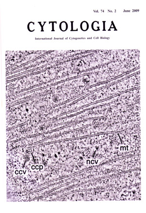 |
|---|---|
| Vol. 74 No.2 June 2009 | |
| Technical note | |
|
|
|
| Quantitative Analysis of Vesicles in the Preprophase Band by Electron Tomography
The preprophase band (PPB) of microtubules (MTs) is an array of plant cortical microtubules, produced during the G2 phase of the cell cycle, that runs beneath the plasma membrane around the nucleus. The mature PPB disappears during prometaphase, but the plasma membrane site demarcated by the PPB corresponds to the ultimate cortical site where the cell plate will fuse at the end of cytokinesis. Little is known about how the plasma membrane becomes modified during PPB formation. Electron tomography is a useful tool for the quantitative 3-D analysis of cytoskeletal and membrane architecture in the cell cortex. To identify the mechanism that could modify the plasma membrane at the PPB site, we have carried out a quantitative analysis of all of the vesicles trafficking to and from the plasma membrane, both within and without of the PPB region, in cells preserved by high pressure freezing. To this end, basal segments of 3-day old onion (Allium cepa L. cv. Highgold Nigou) cotyledons were cut, frozen in a high-pressure freezer, freeze-substituted and embedded in Spurr's resin. 250nm-thick outer tangential sections of epidermal cells were mounted in a tilt-rotate specimen holder and observed in a high-voltage microscope. The images were taken at x12,000 from + 60° to - 60° at 1.5° intervals about 2 orthogonal axes. A tomogram was computed for each set of aligned tilts using the R-weighted back-projection algorithm. A tomographic image of a tangentially sectioned PPB (1.42 nm-thick tomographic slice) in a late prophase cell of onion cotyledon epidermis is shown. The tomogram was displayed and analyzed with Imod, the graphics component of the IMOD software package (http://bi03d.colorado.edu/imod/). Abbreviations: ccp, clathrin-coated pit; ccv, clathrin-coated vesicle; ncv, non-coated vesicle; mt, MT. MTS have a diameter of about 25 nm. The use of electron tomography has enabled us to identify, map, and model the clathrin-coated pits, clathrin-coated vesicles, and MTs of PPB regions with a much higher degree of resolution than is possible with thin sections. We have quantified the distribution of clathrin-coated pits and vesicles, as well as of secretory structures, during PPB formation both in serial thin sections and in electron tomograms, and have determined that the number of clathrin-coated vesicles in the cortical cytoplasm underlying the PPB regions is increased compared to the non-PPB regions. In contrast, the number of secretory events inside and outside of the PPB is essentially the same (see Karahara, I., Suda, J., Tahara, H., Yokota, E., Shimmen, T., Misaki, K., Yonemura, S., Staehelin, A., Mineyuki, Y., Plant J. 2009, 57: 819-831). Based on this finding, we postulate that modification of the plasma membrane at the PPB site involves the removal of a subset of plasma membrane proteins via clathrin-coated vesicles. (Ichirou Karaharal , Jinsuke Sudal, Lucas Andrew Staehelini2, Yoshinobu Mineyuki3, 1Department of Biology, Graduate School of Science and Engineering, University of Toyama, Toyama 930-8555, Japan, 2MCD Biology, University of Colorado, Boulder, CO 80309-0347, USA. 3Department of Life Science, Graduate School of Life Science, University of Hyogo, 2167 Shosha, Himeji, Hyogo 671-2280, Japan ) |
|