| ON THE COVER |
 |
|---|---|
| Vol. 72 No.2 June 2007 | |
| Technical note | |
|
|
|
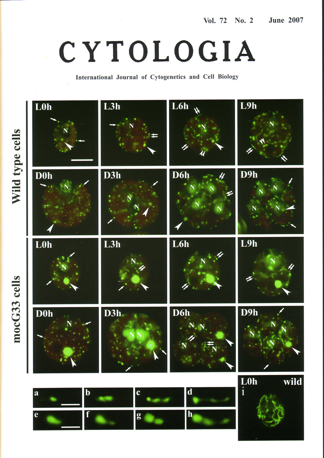
| Morphological changes in the organellar nucleoidsand
mitochondria of living Chlamydomonas reinhardtii Dang were examined during
the cell cycle under conditions of 12:12 light:dark. The nucleoids were
stained with SYBR-Green I , and the mitochondria were stained with 3,3-dihexyloxacarbocyanine
iodide (DiOC6). A mocG33 mutant, which contains one large chloroplast
nucleoid throughout the cell cycle, was used to distinguish between
the mitochondrial and chloroplast nucleoids. Representative sequences of
presumed mitochondrial and chloroplast nucleoid division and the distribution of
newly formed nucleoids were shown. Fluorescence micrographs of cells stained with SYBR Green I (all micrographs except i) or DiOC6 (i). The images of the cells were taken at the onset of the light period (L0h) and at 3h (L3h), 6h (L6h), and 9h (L9h) into the light period, as well as at the beginning of the dark period (D0h) and at 3h (D3h), 6h (D6h), and 9h (D9h) into the dark period. L0h-D0h, an undivided single cell; D3h, a mother cell that contains two daughter cells; D6h, D9h, a mother cell that contains four daughter cells; N, a cell nucleus; arrow, an mitochondrial nucleoid; double arrows, thread-like or bead-like structures of mitochondrial nucleoids; arrowhead, a chloroplast nucleoid, double arrowheads; thread-like or bead-likestructures of cp nucleoids. Scale bar, 5µm. The fluorescent micrographs of the mitochondrial nucleoids (a-d) and chloroplast nucleoids (e-h) are arranged relative to the presumed nucleoid division process. Scale bar,1µm. Living cells were stained directly with SYBR Green I and DiOC6. The final dilution factor of the dye was 1 :10,000. The mixtures of cells were kept in the dark for 5-8 min, after which time 4µl of cell suspension was placed on a glass slide. The cells were observed under blue excitation wavelength (488 nm) with a fluorescence microscope. A red barrier filter was used to reduce red auto-fiuorescence of chlorophyll. Fluorescence photographs were taken with a Nikon digital camera. The contrast of the photographs was increased using the computer software Adobe Photoshop 6.0, when signals were weak. (see Hiramatsu, T., Misumi, O., Kuroiwa, T., and Nakamura, S.-J. Phycol., 42: 1048-l058, 2006). Soichi Nakamura1, Takayoshi Hiramatsu1. Osami Misumi2. and Tsuneyoshi Kuroiwa 2,1Laboratory of Cell and Functional Biology, Faculty of Science, University of the Ryukyus, Nishihara, Okinawa 903-0213 Japan. 2Department of Life Science, College of Science, Rikkyo University. Nishi-Ikebukuro, Tokyo 171-8501 Japan |
|