| ON THE COVER |  |
|---|---|
| Vol. 71 No.2 June 2006 | |
| Technical note | |
|
|
|
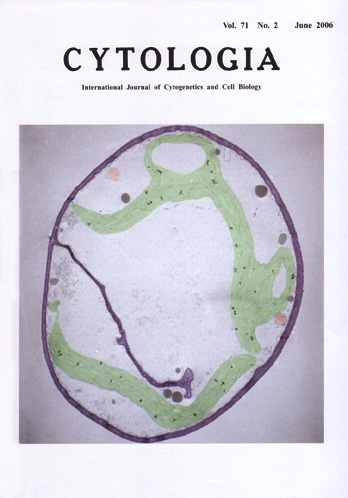
| Electron micrograph of a cell with a new cell wall forming in a Physcomitrella patens MurE knockout transformant The moss Physcomitrella patens has become a model organism for the plant scicnces because it has a simple body plan, it requires simple growth conditions to complete its life cycle, and a gene-targeting technique has been established (Schaefer and Zrÿd 2001 Plant Physiol. 127, 1430: Reski and Cove 2004 Curr. Biol. R261). Chloroplasts in P.patens are easy to observe. In 1997, it was reported that ß-lactam antibiotics, including ampicillin and penicillin, cause a decrease in the number of chloroplasts and the appearance of giant chloroplasts in P. patens (Kasten & Reski 1997 J. Plant Physiol. 150, 137). Subsequently, we demonstrated that fosfomycin and D-cycloserine, which inhibit the peptidoglycan (murein) synthesis pathway at points different from that affected by ß-lactams, also cause the appearance of macrochloroplasts (Katayama et al. 2003 Plant Cell Physiol. 44, 776). A search of the full-length EST library of P. patens identified nine genes related to the bacterial peptidoglycan synthesis pathway in P.patens. Gene disruptant lines for one of these murein genes. PpMurE, were created using a transformation technique. In the PpMurE knockout lines, giant chloroplasts appeared and the number of chloroplasts decreased,similar to the findings in antibiotic-treated cells. These phenotypes suggest a relationship between moss chloroplast division and the remnant bacterial peptidoglycan synthesis pathway. While chloroplasts of normal size do not enter the cell division plane in wild-type cells, the growing cell wall (dark blue) was disturbed by huge chloroplasts (green) in the knockout mutant cell (cover figure). Mitochondria are shown in pink. This electron micrograph suggests that the giant chloroplasts affect cell plate formation. However, time-lapse imaging analysis suggests that the chloroplasts divide during cell division, although the detailed mechanism of chloroplast division during cell division is unclear. Time-lapse observations may help to answer the question of why we do not observe cells without chloroplasts in the PpMurE disruptant lines. (See Machida et al. 2006 Proc. Natl. Acad. Sci. USA 103, 6753.) (Katsuaki Takechi, Hiroshi Sato and Hiroyoshi Takano, Graduate School of Science and Technology, Kumamoto University, Kumamoto 860-8555, Japan) |
|