| ON THE COVER | 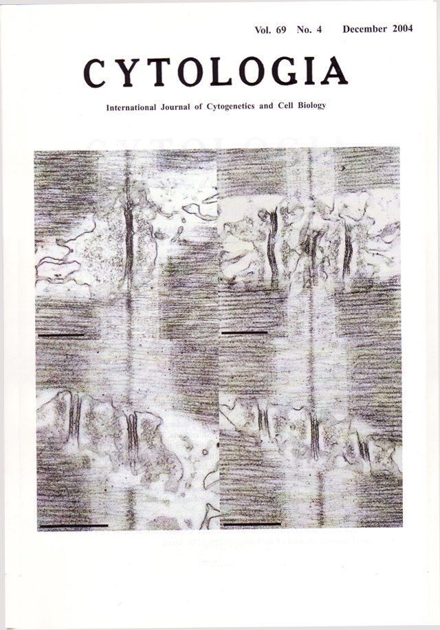 |
|---|---|
| Vol. 69 No.4 December 2004 | |
| Technical note | |
|
|
|
| Various T
tubule-SR contacts in the fish swim-bladder muscle: two different types of triads, pentads and heptads. In skeletalmuscle fibers, the influence of action potential at the surface membrane spreads inwards along the membrane of transverse tubule (T tubule), and causes Ca2+ release from the terminal cisternae of sarcoplasmic reticulum (SR). Then, under the electron microscope, it has been recognized that a T tubule forms generally a triadic contact (triad) with a pair of terminal cisternae of SR, the T tubule-SR contact. Based on the sarcomeric location of contacts, two different types of triads have been established. One is a 'Z-type triad', which is located around the level of the Z-band in each sarcomere (upper left figure, scale bar, 0.5µm). The other is a 'AI-type triad', which is located around the level of boundary between the A- and I-bands in each sarcomere (upper right figure, scale bar, 0.5µm). It has been believed that every kind of muscle fiber contains only one type of triadic contact. Recently, we found that two different types of triadic contacts were present within the single swim-bladder muscle fiber of a scorpionfish, Sebastiscus marmoratus (upper figures). They were regularly distributed within the single fiber; the Z-type triads were located in the two end regions of the fiber, while the Al-type triads were found in the middle region of the fiber. Furthermore, between the end and middle regions of the fiber, we found also two types of complex T tubule-SR contacts. They are 'pentad' composed of a pair of T tubules and three SR elements (lower left figure, scale bar, 0.5µm, and newly named 'heptads' composed of three T tubules and four SR elements (lower right figure, scale bar, 0.5µm). The structural and functional relation among two types of triads, pentads and heptads found in the same fiber has been discussed, in connection with the prominent function of this muscle, producing audible sounds by its rapid contraction-relaxation cycle (See Suzuki S., Nagayoshi H., Ishino K., Hino N., and Sugi H. 2003: J. Electron Microsc.. 52: 337-347). (Suechika Suzuki, Department of Biological Sciences, School of Science, Kanagawa University, Hiratsuka-City, Kanagawa 259-1293, Japan) |
|