| ON THE COVER | 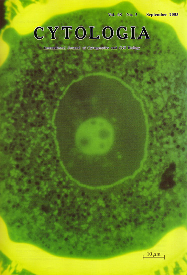 |
|---|---|
| Vol. 68 No.3 September 2003 | |
| Technical note | |
|
|
|
| Mitochondria cover up the nuclear surface
at unicellular stage in microspores of Pharbitis nil. Fluorescence microscopic observation of the thin sections of pollen grains, which were embedded in Technovit 7100 resin and stained with 3, 3'-dihexyloxacarbocyanine iodide (DiOC6), was found to allow observation of mitochondria at high resolution. Thin sections (about 1 µm thick) were stained with 100µg/ml DiOC6 in ethanol, washed in distilled water, then they were observed using a fluorescence microscopy. DiOC6 clearly visualized the mitochondria as uniform spherical object. The technique is effective for examining the distribution of mitochondria throughout large cells, such as P. nil pollen grains ( more than 100µm in diameter on a flowering day). We observed the changes in the distribution of mitochondria in microspores/pollen grains of P. nil during male gametogenesis. During the process, mitochondria exhibited characteristic localization twice; subpopulation of mitochondria covered up the surface of microspore- and vegetative-nucleus 2 days before and after pollen mitosis one (PMI), respectively (9 days and 5 days before flowering). The photograph shows positioning of mitochondria 2 days before PMI. The characteristic localization of mitochondria in P. nil pollen may serve as an excellent model to analyze mechanisms that control mitochondrial positioning within a cell and interactions between mitochondria and nuclei (see Nagata, N., Saito, C., Sakai, A., Kuroiwa, H., Kuroiwa, T. 2000: Protoplasma, 213: 74-82). (Noriko Nagata, Department of Chemical Biological Sciences, Faculty of Science, Japan Women's University, Mejirodai, Bunkyo-ku, Tokyo, 112-8681, Japan) |
|