| ON THE COVER | 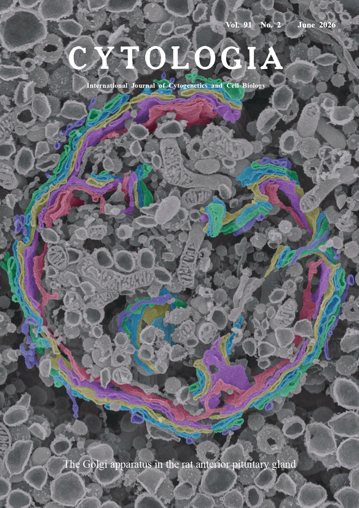 |
||
|---|---|---|---|
| Vol. 91 No.2 June 2026 | |||
| Technical Note | |||
|
|
|||
| Three-dimensional ultrastructure of the Golgi apparatus visualized by scanning
electron microscopy Daisuke Koga* Department of Microscopic Anatomy and Cell Biology, Asahikawa Medical University, Asahikawa 078–8510, Japan
The Golgi apparatus is a cellular organelle first discovered by Camillo Golgi in 1898. Since then, extensive morphological studies of the Golgi apparatus have been conducted primarily using light microscopy and transmission electron microscopy. Nevertheless, the precise three-dimensional (3D) ultrastructure of this organelleremains unclear because its complex spatial organization cannot be adequately interpreted from single sections. To address this issue, we applied the osmium maceration method, a unique technique that enables direct visualization of the surface structures of membranous organelles using scanning electron microscopy, to elucidate the morphological features of the Golgi apparatus in three dimensions (Koga et al. 2024). Using this macerationmethod, we examined the Golgi apparatus in mammalian cells and demonstrated that its 3D ultrastructure varies depending on the cell type (Koga et al. 2025). The cover figure shows a scanning electron microscopy image of the Golgi apparatus in a rat pituitary gonadotrope prepared using the osmium maceration method. The animal experiments were approved by the Committee of Ethics on Animal Experiments at Asahikawa Medical University and were carried out in accordance with the Asahikawa Medical University Animal Experimentation Regulations (Approval No. R7-103). An adult male rat, 10 weeks of age, was used. Briefly, after deep anesthesia, the animal was perfused with a mixture of 0.5% paraformaldehyde and 0.5% glutaraldehyde. The pituitary gland was excised from the animal and immersed in 1% osmium tetroxide (OsO4) for 1 h. After the tissue was immersed in 25% and 50% dimethyl sulfoxide for 30 min each, it was frozen on a metal plate pre-cooled with liquid nitrogen and freeze-fractured into two pieces using a screwdriver and hammer. The pieces were thawed in 50% dimethyl sulfoxide. For cell maceration, the tissue blocks were immersed in 0.1% OsO4 for 72 h at 20°C. This step is essential for removing cytoplasmic soluble proteins from freeze-cracked cell surfaces. The macerated specimens were fixed with 1% OsO4, treated with 1% tannic acid, and re-immersed in 1% OsO4. The samples were then dehydrated through a graded ethanol series and dried in a critical-point dryer. The dried specimens were mounted onto an aluminum base, coated with platinumpalladium, and observed under a field-emission scanning electron microscope (Regulus, Hitachi). Gonadotropes contain a spherical Golgi apparatus (Koga et al. 2017). The endoplasmic reticulum–Golgi intermediate compartment, cis cisterna, medial cisternae, trans cisterna, and trans-Golgi network are pseudocolored blue, green, light blue, yellow, purple, and red, respectively. The cis and trans faces are located on the outer and inner sides of the sphere, respectively. The Golgi stack consists of several sheet-like cisternae that accumulate in a concentrated manner. The cisternae within the Golgi stacks are intricately interwoven in some regions of the spherical structure. As shown in the cover figure, the osmium maceration method provides detailed 3D visualization of the Golgi apparatus, unlike transmission electron microscopy of single ultrathin sections. It also offers the potential for 3D structural analysis of organelles, including not only the Golgi apparatus but also the endoplasmic reticulum and mitochondria.
Koga, D., Kusumi, S., Yagi, H., and Kato, K. 2024. Three-dimensional analysis of the intracellular architecture by scanning electron microscopy. Microscopy (Oxf.) 73: 215–225. Koga, D., Morinaga, R., and Kusumi, S. 2025. Three-dimensional ultrastructure of the golgi apparatus in vivo: Scanning electronmicroscopy of osmium-macerated mammalian cells. Subcell.Biochem. 110: 1–34. * Corresponding author, e-mail: daisukek@asahikawa-med.ac.jp DOI: 10.1508/cytologia.91.79 |
|||
 Back Archive list English Top Japanese Top Back Archive list English Top Japanese Top |
|||