| ON THE COVER | 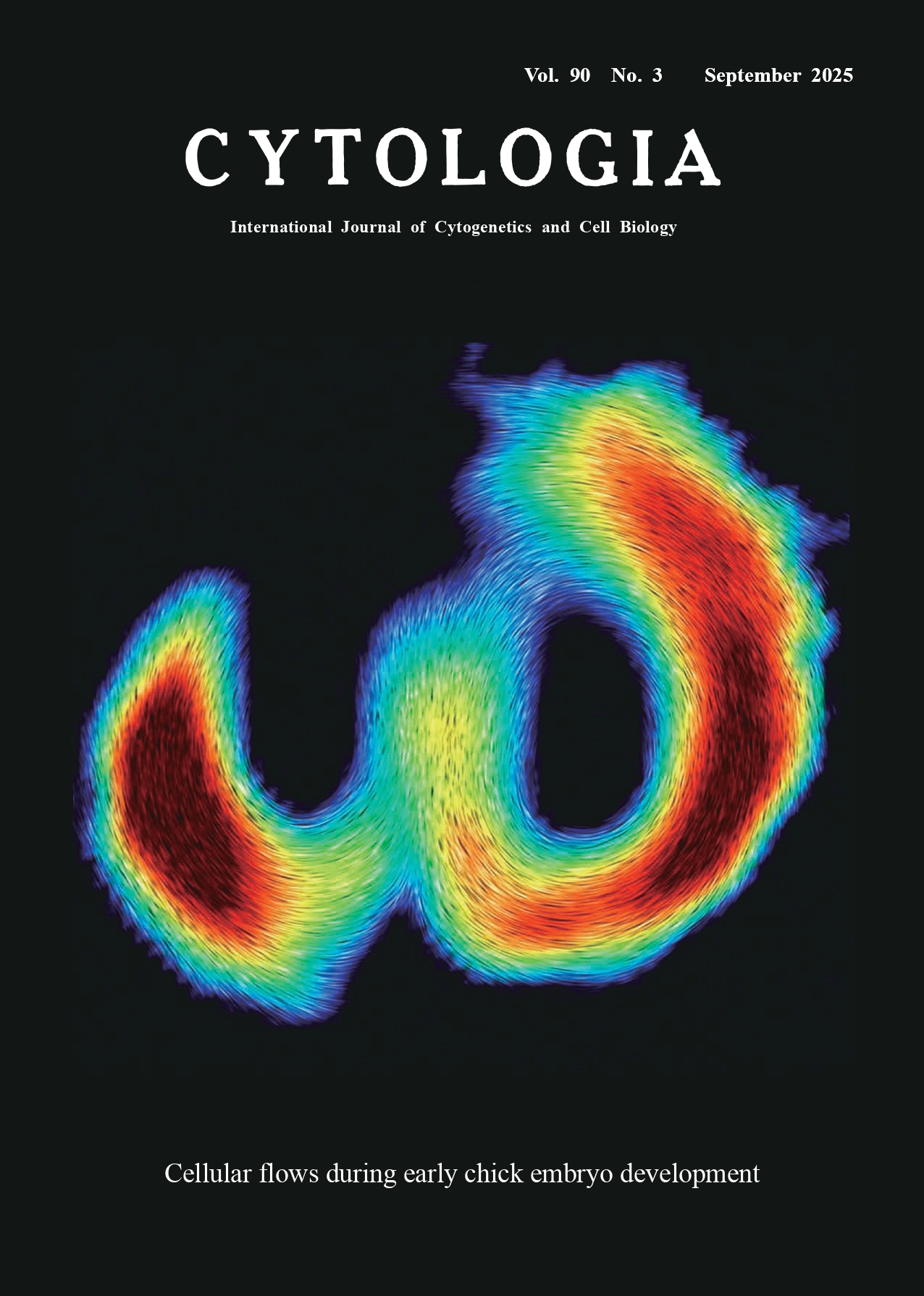 |
||
|---|---|---|---|
| Vol. 90 No.3 September 2025 | |||
| Technical Note | |||
|
|
|||
| Live-imaging with quantitative analysis of cellular flows to visualize
left-right asymmetry during early chick embryo development Rieko Asai1,2* Shubham Sinha3, Vivek N. Prakash3,4,5*, and Takashi Mikawa1* 1 Cardiovascular Research Institute, University of California, San Francisco, San Francisco, California 94158, USA 2 Kumamoto University, IRCMS, Kumamoto, 860–0811, Japan 3Department of Physics, University of Miami, Coral Gables, Florida 33146, USA 4Department of Biology, University of Miami, Coral Gables, Florida 33146, USA 5Department of Marine Biology and Ecology, University of Miami, Miami, Florida 33149, USA
The cover image generated by Shubham Sinha, Drs. Santhan Chandragiri and Vivek Prakash, which was originally from our latest study (Asai et al. 2025) illustrates the LR asymmetry in bilateral cellular flows during primitive streak formation in a chick embryo. Cell speed is color-coded in the image, with red representing areas of maximum speed and blue representing areas of minimal speed. The lines or streaks represent the velocity vector field, and were calculated using the line integral convolution (LIC) method of flow visualization. Since imaging was performed dorsally using an inverted microscope, the right side of the image corresponds to the right side of the embryo. Thus, this speed heatmap indicates that the area and the speed of the cell movements in the right side was larger compared to the left side of the embryo. Here, we briefly describe the materials and methods underlying this image. An unincubated chick embryo, known as an “embryonic disc” or “blastoderm”, was isolated from the vitelline membrane and yolk in a Tyrode’s solution and transfected with fluorescent-conjugated control morpholino by electroporation. The fluorescently tagged embryo was then transferred onto a new vitelline membrane, which had been stretched over a glass ring placed on albumin in a 35-mm dish. The dish containing the ex ovo cultured embryo was placed on the stage of an inverted microscope (Nikon Ti inverted fluorescent microscope with CSU-W1 scanner), which was enclosed in a dark hood equipped with a heating system maintained at 37°C. Liveimaging of the embryo was performed for approximately 10 h prior to and during primitive streak formation. The recorded sequence of images was post-processed using PIV (PIVlab package in MATLAB) to quantify the velocity and vorticity of the cellular flows. Despite the presence of bilateral cellular flows, the underlying mechanisms of their LR-asymmetry remain unclear. Classical methods of in situ hybridization for laterality genes (e.g., Lefty, Shh, and Nodal) did not detect LR-asymmetric expression during the same developmental stages as the cellular flows (Asai et al. 2025). Future state-of-the-art analyses may help elucidate the mechanisms of LR asymmetry in the cellular flows and their contribution to LR asymmetric body patterning.
Asai, R., Prakash, V. N., Sinha, S., Prakash, M., and Mikawa, T. 2024. Coupling and uncoupling of midline morphogenesis and cell flow in amniote gastrulation. eLife 12: RP89948. Asai, R., Sinha, S., Prakash, V. N., and Mikawa, T. 2025. Bilateral cellular flows display asymmetry prior to left–right organizer formation in amniote gastrulation. Proc. Natl. Acad. Sci. U.S.A. 122: e2414860122. * Corresponding author, e-mail: rasai@kumamoto-u.ac.jp; vprakash@miami.edu; takashi.mikawa@ucsf.edu DOI: 10.1508/cytologia.90.145 |
|||