ON  THE  COVER   
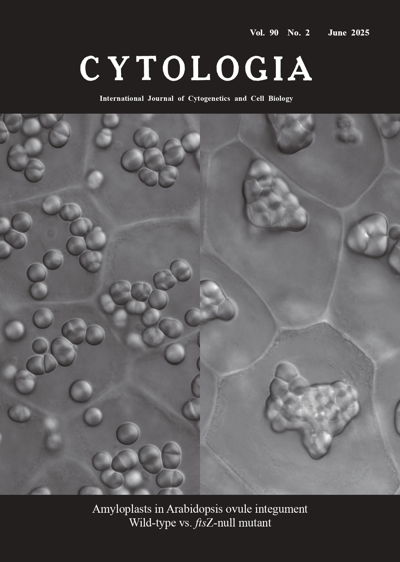
 Vol. 90   No.2  June  2025
 Technical Note 

Amyloplast imaging system in Arabidopsis ovule integument

Makoto T. Fujiwara1* and Ryuuichi D. Itoh2

1 Department of Materials and Life Sciences, Faculty of Science and Technology, Sophia University, Tokyo 102–8554, Japan
2 Department of Chemistry, Biology and Marine Science, Faculty of Science, University of the Ryukyus, Senbaru 1, Nishihara, Okinawa 903–0213, Japan

Received April 10, 2025; accepted April 23, 2025
    Plastids are unique organelles that exhibit remarkable morphological differentiation in seed plants. Undifferentiated proplastids in meristematic tissues undergo active proliferation and differentiation, giving rise to photosynthetic chloroplasts in leaves, carotenoidaccumulating chromoplasts in fruits and flowers, and non-pigmented leucoplasts in roots. Amyloplasts represent a non-photosynthetic type of plastid that accumulates starch in seeds, fruits, stems, and roots. Plastids replicate by division of preexisting organelles through the constriction of the envelope membranes. Research into the molecular mechanisms of plastid replication has largely been conducted using chloroplasts. The replication mechanism in amyloplasts remains unclear, owing to the lack of a suitable live-analysis system. Recently, we proposed that the outer ovule integument of Arabidopsis thaliana could facilitate live imaging of amyloplast replication. Using this system, we demonstrated that the regulatory basis of amyloplast replication differs from that of chloroplasts (Fujiwara et al. 2024).    The cover shows differential interference contrast (DIC) images of the outer integument cells of mature ovules (Phase III, as defined in Fujiwara et al. 2024) from A. thaliana wild-type plants (Columbia accession; left panel) and the ftsZ1-1 ftsZ2-1 ftsZ2-2 triple mutant (ftsZnull; a gift from Prof. K.W. Osteryoung; Schmitz et al. 2009; right panel). In brief, siliques of 5-to-6-week-old plants were dissected on a glass slide under an MZ10 F stereomicroscope (Leica Microsystems, Heidelberg, Germany). A septum-ovule association was extracted by peeling off one or both valves and transferred to a drop of sterilized water on a second glass slide. A coverslip was carefully mounted. High resolution imaging of amyloplasts in the outer integument cells of ovules was performed using an IX71 inverted microscope (Olympus, Tokyo, Japan) with a UPlanSApo 60×/1.20 W water-immersion objective lens (Olympus) and an ORCA-flash2.8 camera (Hamamatsu Photonics, Hamamatsu, Japan). Detailed procedures and technical tips are described in our recent publication (Fujiwara et al. 2025).
   Our imaging technique revealed distinct amyloplast morphologies in the ovule integument of wild-type and ftsZ-null mutants of A. thaliana. Wild-type cells contained round to ellipsoidal amyloplasts of relatively homogeneous size. By contrast, ftsZ-null mutant amyloplasts displayed severe morphological anomalies, characterized by irregular, enlarged amyloplasts with excessive stromule formation (Fujiwara et al. 2024). The number of amyloplasts per cell was low (1–3 in Phase III) in the ftsZ-null mutant compared to the wild type (9–15 amyloplasts in Phase III). The mutant amyloplasts also exhibited aberrant starch granule distribution, as starch granules were often localized within stromules in mutant amyloplasts but occupied most of the organelle volume in wild-type amyloplasts. These findings underscored the critical role of FtsZ in mediating division and maintaining structural integrity of amyloplasts. The absence of FtsZ disrupted envelope fission and led to unregulated amyloplast enlargement, while also impairing starch compartmentalization, highlighting its indispensability for normal amyloplast biogenesis and replication in A. thaliana.
    We hope that the system for imaging amyloplasts in A. thaliana ovule integument introduced here will serve as a foundation for future advances in our understanding of amyloplast biogenesis.
References
Fujiwara, M. T., Arakawa, R., Abe, T., and Itoh, R. D. 2025. Using a live analysis system to study amyloplast replication in Arabidopsis ovule integuments. Bio Protoc. 15: e5333.

Fujiwara, M. T., Yoshioka, Y., Kazama, Y., Hirano, T., Niwa, Y., Moriyama, T., Sato, N., Abe, T., Yoshida, S., and Itoh, R. D. 2024. Principles of amyloplast replication in the ovule integuments of Arabidopsis thaliana. Plant Physiol. 196: 137–152.

Schmitz, A. J., Glynn, J. M., Olson, B. J. S. C., Stokes, K. D., and Osteryoung, K. W. 2009. Arabidopsis FtsZ2-1 and FtsZ2-2 are functionally redundant, but FtsZ-based plastid division is not essential for chloroplast partitioning or plant growth and development. Mol. Plant 2: 1211–1222.

* Corresponding author, e-mail: mtf1@mac.com
DOI:
10.1508/cytologia.90.77


Back          Archive list         English Top      Japanese Top