| ON THE COVER | 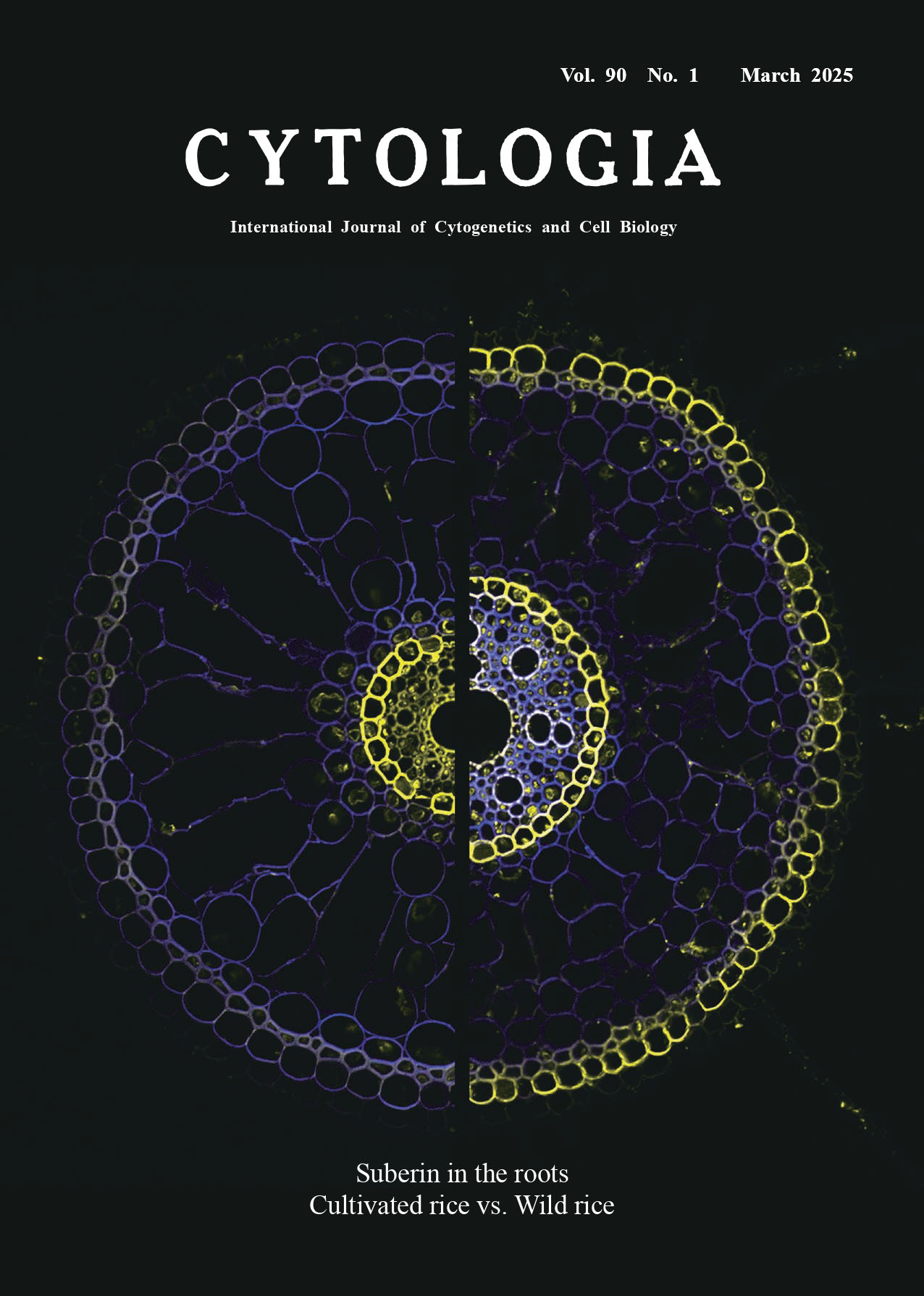 |
||
|---|---|---|---|
| Vol. 90 No.1 March 2025 | |||
| Technical Note | |||
|
|
|||
| High resolution detection of suberized cells in root cross-sections using Z-stack images
Masato Ejiri1,2 and Katsuhiro Shiono2* 1 Research and Development Department, Fitech Co., Ltd, 3–101 Akita, Oguchi, Niwa, Aichi, 480–0132, Japan 2 Graduate School of Bioscience and Biotechnology, Fukui Prefectural University, 4–1–1 Matsuoka-Kenjojima, Eiheiji, Fukui 910–1195, Japan
Suberin is a hydrophobic biopolymer that forms a diffusion barrier at the periphery of plant cells, helping them resist biotic and abiotic stresses. In plant roots, suberin accumulates around endodermal and exodermal cells and is crucial for controlling water, nutrient, and gas transport via extracellular spaces (apoplast). In the roots of wetland plants, suberized exodermis (the outset cell layer of the cortex) plays the role of a diffusion barrier against oxygen loss, enhancing long-distance oxygen transport and preventing the influx of soil phytotoxins from the waterlogged soils. This barrier at the exodermis, called a radial oxygen loss (ROL) barrier, is essential to acclimate plants to waterlogged conditions. ROL barriers can be classified as inducible or constitutive (Shiono and Nakazono 2024). In some wetland species, including rice, an ROL barrier is induced in roots when the plants are grown in waterlogged soil or hydroponic systems that mimic waterlogging, whereas the roots remain permeable to oxygen when grown in well-drained soil or aerated hydroponics. ROL barriers form constitutively in several other wetland species, including wild rice (Oryza glumaepatula), which develops even in the absence of waterlogging. The ability to form either an inducible or constitutive barrier is considered an acclimation to waterlogged conditions. The cover figure presents confocal microscopy images of suberin deposits (yellow) at the exodermis and the endodermis in root cross-sections of cultivated rice (left) and O. glumaepatula (right). The suberin is stained with Fluorol Yellow 088 (FY) as previously described (Ejiri and Shiono 2023). Suberin lamellae stained with FY are visualized as a yellow fluorescence excited by ultraviolet light. Cell walls were detected as blue autofluorescence excited by ultraviolet light. In this note, capturing suberin distribution in a larger root cross-section (e.g., rice roots with 1 mm in diameter) can be challenging due to variations in the cross-section thickness. To obtain high resolution images of suberin deposits in root cross-sections, we used maximum intensity projection (MIP), in which the voxel with the highest attenuation value on every focal depth in a Z-stack is projected onto a 2D image. In this study, the basal portions (75 to 85 mm from the root apex) of a seminal root (95 to 105 mm length) were embedded in 6% (w/v) agar, and 70 μm sections were made using a vibrating microtome (Leica VT1200S, Leica Biosystems, Wetzlar, Germany). Suberin lamellae were detected with 0.01% (w/v) FY in polyethylene glycol 400. We captured images with a confocal laser scanning microscope (LSM900, Carl Zeiss, Oberkochen, Germany) with the following settings to obtain specific fluorescence signals: FY, excitation laser: 488 nm, collection bandwidth: 520–580 nm; Autofluorescence, excitation laser: 405 nm, collection bandwidth: 400–480 nm. Three different images in a cross-section were captured in the 3.5–5.0 μm range along the Z-axis for a single cross-section. The Z-stack images were then reconstructed using the function of MIP. The ratio of suberized cells at the exodermis was quantified with image analysis software (Fiji version 2.1.0). Under aerated conditions, we found that the rate of suberized exodermis was 1% in cultivated rice and 65% in O. glumaepatula, respectively (Ejiri and Shiono 2023). O. glumaepatula has an AA genome (a reservoir of favorable alleles that can be used to improve rice crops), which allows for potential crossing with cultivated rice that does not form a constitutive barrier. Currently, we are jmapping constitutive ROL barrier-related quantitative trait loci in introgression lines of O. glumaepatula to identify the genes that regulate the formation of constitutive ROL barriers.
Shiono, K. and Nakazono, M. 2024. Development and regulation of a radial oxygen loss barrier to acclimate to anaerobic conditions. In: Sakagami, J. and Nakazono, M. (eds.). Responses of Plants to Soil Flooding. Springer Nature Singapore, Singapore. pp. 119–138. * Corresponding author, e-mail: shionok@fpu.ac.jp DOI: 10.1508/cytologia.90.1 |
|||