| ON THE COVER |  |
||
|---|---|---|---|
| Vol. 89 No.3 September 2024 | |||
| Technical Note | |||
|
|
|||
Visualization of the shell structure of the testate amoeba Paulinella micropora by FE-SEM Mami Nomura* Faculty of Science, Yamagata University, 1–4–12 Kojirakawa-machi, Yamagata, Yamagata 990–8560, Japan
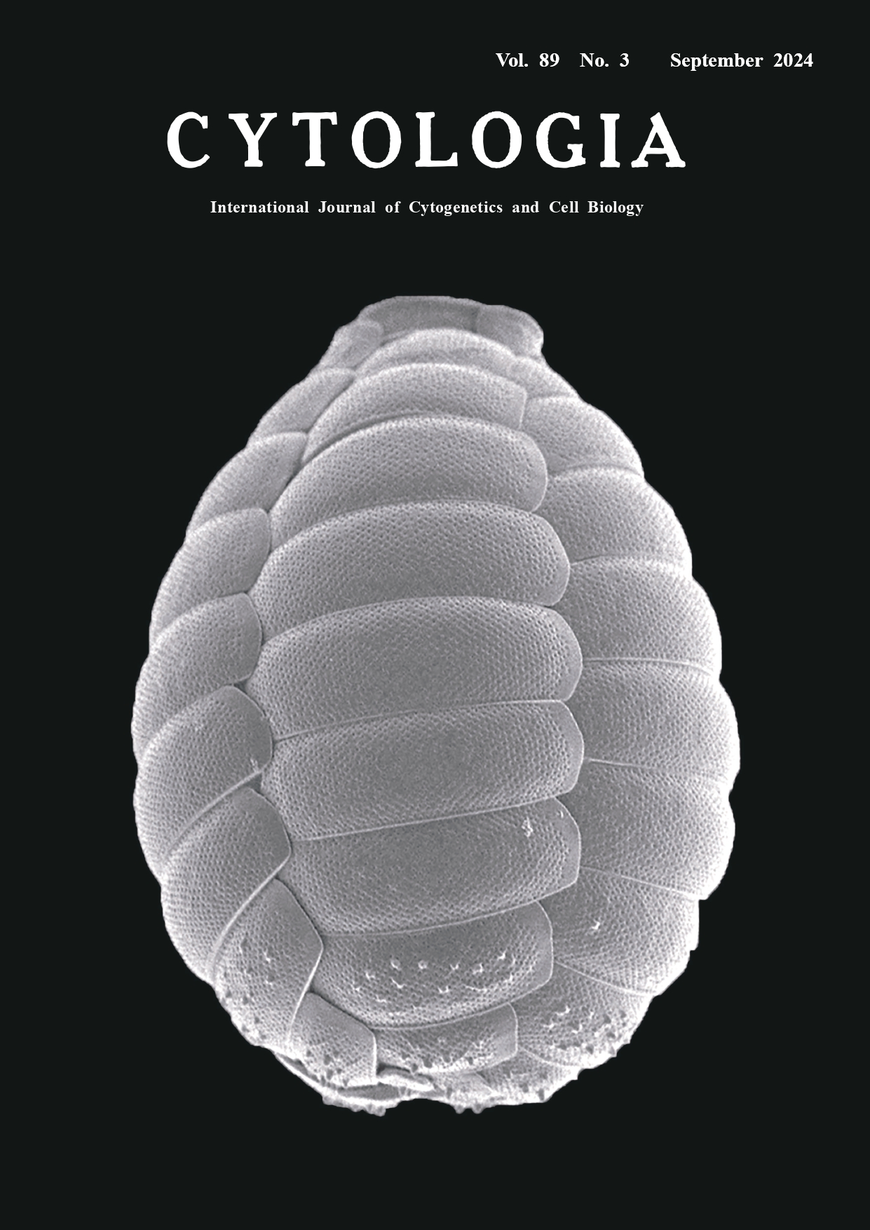
Amoebae belonging to the Euglyphida in Rhizaria have shells, which are mostly constructed by self-secreted siliceous scales. Photosynthetic Paulinella species, which belong to the Euglyphida, are testate amoebae with photosynthetic organelles (chromatophores) acquired by independent primary endosymbiosis with a cyanobacterium distinct from chloroplasts (Nowack 2014). The cover figure of this issue shows a scanning electron microscope (SEM) image of a photosynthetic Paulinella micropora shell. This shell is composed of approximately 50 scales which arranged five sets of scale rows with 10 scales differ in size and shape along the longitudinal axis of the shell. On the other hand, it has been difficult to understand the three-dimensional (3D) orientation of the scales in the shell in detail using transmission and/or scanning electron microscopes, which basically provide only two-dimensional images. Recently, 3D reconstruction analysis using focused ion beam scanning electron microscopy (FIB-SEM) has revealed how individual scales are arranged in three dimensions, down to their positional relationships (Nomura et al. 2023). It has also been shown that the scales composing the shell all have different shapes. During the process of shell construction, it was observed that they skillfully manipulate the extracellular scales using a specialized thick pseudopodium for shell construction (Nomura et al. 2014; Nomura and Ishida 2016). The shell on the SEM image was obtained from a P. micropora strain NIES-4060. To prepare the SEM observation, a fresh culture of strain NIES-4060 was dropped onto a 0.2 μm Isopore™ hydrophilic polycarbonate membrane (Merck Millipore, MA, USA) and incubated for a few minutes to fix the cells to the membrane. Prior to mounting the cells, the membrane, cut to the size of the SEM stub, was placed on filter paper and hydrophilized by dropping deionized water on it. The samples on the membrane were then washed with a drop of deionized water to wash out the culture medium components. The samples on the membrane were well dried in air and mounted on a 12.5×10 mm diameter SEM stub (EM Japan, Tokyo, Japan) with carbon paste Pelco colloidal graphite (Ted Pella Inc., Redding, CA, USA) or carbon double-sided conductive carbon tape (Nisshin EM Co., Ltd., Tokyo, Japan). When carbon paste was used, the samples were air dried for at least one day until the organic solvent was completely removed. Well-dried samples were coated with osmium tetroxide to a thickness of approximately 5 nm using an osmium coater Tennant 20 (Meiwafosis Co., Ltd., Tokyo, Japan) and observed by Field emission scanning electron microscopy (FE-SEM) JSM-7600F (JEOL Ltd., Tokyo, Japan).
Nowack, E. C. 2014. Paulinella chromatophora-rethinking the transition from endosymbiont to organelle. Acta Soc. Bot. Pol. 83: 387–397. Nomura, M. and Ishida, K. I. 2016. Fine-structural observations on siliceous scale production and shell assembly in the testate amoeba Paulinella chromatophora. rotist 167: 303–318. Nomura, M., Nakayama, T., and Ishida, K. I. 2014. Detailed process of shell construction in the photosynthetic testate amoeba Paulinella chromatophora (Euglyphid, hizaria). J. Eukaryot. Microbiol. 61: 317–321. Nomura, M., Ohta, K., Nishigami, Y., Nakayama, T., Nakamura, K. I., Tadakuma, K., and Galipon, J. 2023. Three-dimensional architecture and assembly mechanism of the egg-shaped shell in testate amoeba Paulinella micropora. Front. Cell Dev. Biol. 11: 1232685. * Corresponding author, e-mail: mami_nomura_4p@sci.kj.yamagatau.ac.jp DOI: 10.1508/cytologia.89.179 |
|||