| ON THE COVER | 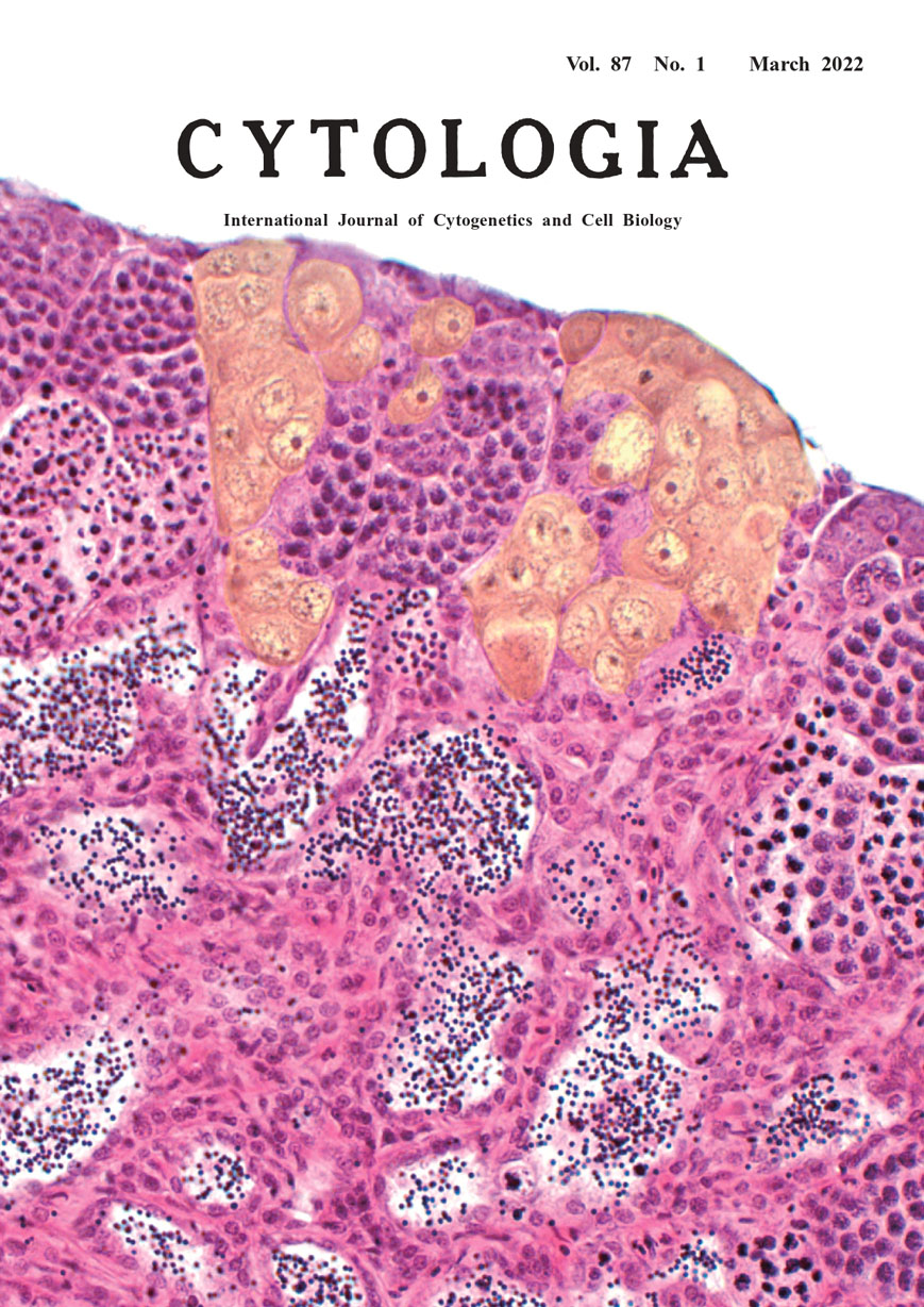 |
||
|---|---|---|---|
| Vol. 87 No.1 March 2022 | |||
| Technical Note | |||
|
|
|||
Testis-ova Induction by Microbeam Irradiation in P53-Deficient Medaka Testis Kento Nagata1,*, Takako Yasuda1, Michiyo Suzuki2, Tomoo Funayama2,Hiroshi Mitani1 and Shoji Oda1 1 Department of Integrated Biosciences, Graduate School of Frontier Sciences, The University of Tokyo, 5–1–5 Kashiwanoha, Kashiwa, Chiba 277–8562, Japan 2 Takasaki Advanced Radiation Research Institute, Quantum Beam Science Research Directorate, National Institutes for Quantum Science and Technology (QST), 1233 Watanuki, Takasaki, Gunma 370–1292, Japan
The Japanese medaka, Oryzias latipes, is a small teleost fish that has been used as a model organism to investigate the biological effects of radiation in vertebrates. A testis-ova can be differentiated in medaka testis lacking the p53 gene when exposed to gamma-ray radiation (Yasu da et al. 2012). Estrogen administration has also been reported to result in testis-ova differentiation with sexual bipotentiality, including the production of type A spermatogonia (Ga) (Shibata and Hamaguchi 1988). On the other hand, an increase in serum estrogen is observed in irradiated mice (Suman et al. 2012). However, it is not clear whether the differentiation of medaka testis-ova by radiation is mediated by increased serum estrogen levels. We have previously established a whole-body protocol using broad-beam irradiation to adult medaka (Nagata et al. 2016). Here, we adopted the microbeam irradiation method to verify whether abnormal differentiation of spermatogonia could occur locally in p53-deficient fish. We established experimental procedures to irradiate medaka to a depth of approximately 2.2 mm from the body surface using the heavy-ion beam system of QSTTakasaki (Funayama 2019). The testis is located in the body cavity along the swim bladder. Based on the position of the anus, microbeam irradiation was performed at five locations along the body cavity with a width of 2–3 mm. The cover image shows a cluster of testis-ova cells with enlarged nucleoplasm and condensed nucleoli in a cyst of Ga presumably located in the path of the microbeam (250 μm wide, 320 MeV, 2 Gy) at 7 days after the targeted irradiation by carbon ions (highlighted in yellow). A cluster of testis-ova cells was also found on the opposite side of the testis. In contrast, only normal spermatogonia were present in other Ga cysts surrounding the testis-ova cluster as in the non-irradiated control testis. These results indicate that testis-ova could be induced from Ga exposed to carbon-ion beams, as with whole-body gamma ray irradiation. This suggests that radiation-inducible hormonal signaling might not be involved in testis-ova formation. Because of their small size, cells in the inner tissues of medaka can be irradiated with microbeams, which have a shallow depth of penetration from the surface. The beam size can be used to distinguish between damaged and surrounding cells. This technique could be used to understand the effects of radiotherapy and chemotherapy on reproductive stem cell differentiation. Thus, the molecular machineries of testis-ova induction might be elucidated by a combination of the targeted irradiation established here and spatial transcriptomics studies.
Funayama, T. 2019. Heavy-ion microbeam for biological science: Development of system and utilization for biological experiments in QST-Takasaki. Quantum Beam Sci. 3: 13. Nagata, K. et al. 2016. In vivo 3D analysis of systemic effects after local heavy-ion beam irradiation in an animal model. Sci. Rep. 6: 28691. Shibata, N. and Hamaguchi, S. 1988. Evidence for the sexual bipotentiality of spermatogonia in the fish, Oryzias latipes. J. Exp. Zool. 245: 71–77. Suman, S., Johnson, M. D., Fornace, A. J. Jr. and Datta, K. 2012. Expo sure to ionizing radiation causes long-term increase in serum estradiol and activation of PI3K-Akt signaling pathway in mouse mammary gland. Int. J. Radiat. Oncol. Biol. Phys. 84: 500–507. Yasuda, T., Oda, S., Li, Z., Kimori, Y., Kamei, Y., Ishikawa, T., Todo, T. and Mitani, H. 2012. Gamma-ray irradiation promotes premature meiosis of spontaneously differentiating testis-ova in the testis of p53-deficient medaka (Oryzias latipes). Cell Death Dis. 3: e395. * Corresponding author, e-mail: nagata.kento@qst.go.jp DOI:10.1508/cytologia.87.1 |
|||