| ON THE COVER | 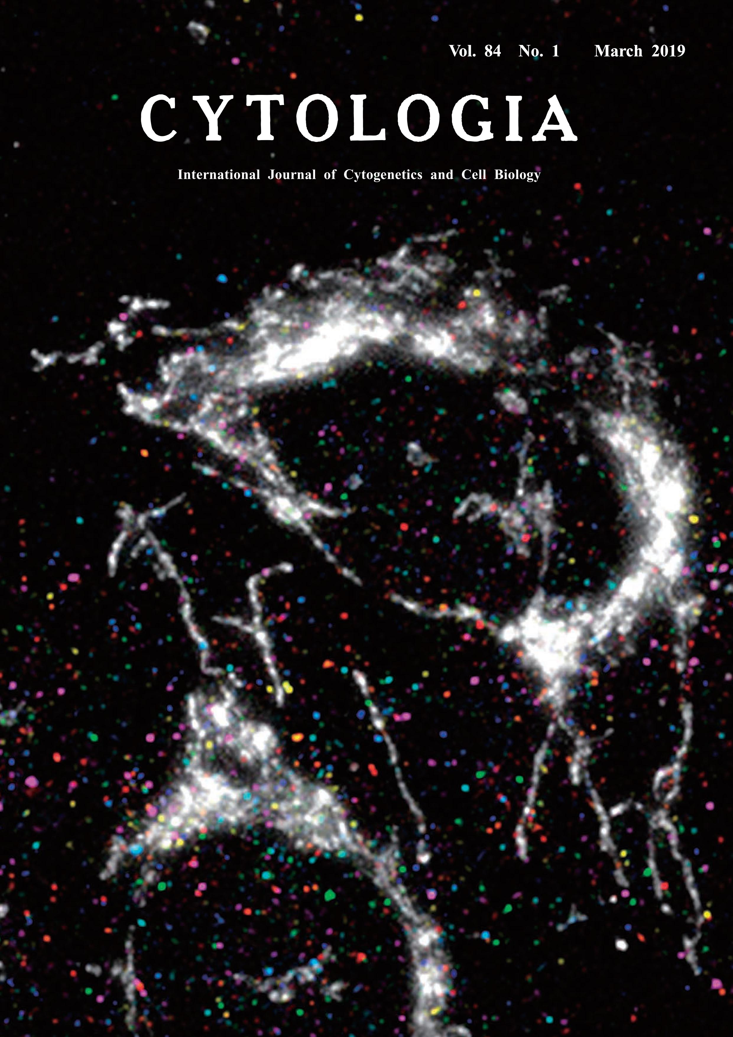 |
|
|---|---|---|
| Vol. 84 No.1 March 2019 | ||
| Technical Note | ||
|
|
||
Simultaneous Measurements of RNA Molecules Transcribed from Mitochondrial Division or Fusion Genes in Single Cells Yamato Yoshida1* and Yuichi Taniguchi2* 1 Department of Science, College of Science, Ibaraki University, 2–1–1 Bunkyo, Mito, Ibaraki 310–8512, Japan 2 Laboratory for Cell Systems Control, RIKEN Center for Biosystems Dynamics Research, 6–2–3 Furuedai, Suita, Osaka 565–0874, Japan Received December 29, 2018; accepted January 18, 2019 Mitochondria are descended from an endosymbiotic bacterial ancestor (Gray 1992). Due to the evolutional origin, their continuity maintains by division of preexisting mitochondria (Yoshida and Mogi 2018). In addition, mitochondria in higher organisms can perform not only division but also fusion. Thus, the intracellular distribution of mitochondria is a highly dynamic process in which mitochondria constantly divide and fuse. Several genes which are required for mitochondrial division and fusion have been characterized in human, such as DRP1, FIS1, MFF, and MIEF1/2 for division and MFN1/2 and OPA1 for fusion. The loss of function of mitochondrial division genes causes excess fusion of mitochondria, leading to the presence of long, interconnected mitochondria. By contrast, the loss of function of mitochondrial fusion genes leads to excess divisions, with extensive fragmentation of the mitochondria. However, it is still unclear how the mitochondrial division and fusion processes are coordinately regulated in the normal cell. To quantify RNA expression levels in an individual cell, single-molecule fluorescence in situ hybridization (smFISH) has been developed to identify the copy number and spatial distribution of an RNA molecule of interest (Raj et al. 2008). Here, we demonstrated microscopic observation using smFISH and immunostaining to simultaneously measure RNA molecules transcribed from mitochondrial or fusion genes and the number of mitochondria in an individual cell (Yoshida and Taniguchi 2019). For sequential smFISH imaging, U2-OS cells were cultured on 35 mm φ glass bottom dishes (Matsunami). The cultured cells were fixed in 4% paraformaldehyde and washed with 1×PBS. Each RNA species transcribed from mitochondrial division or fusion genes was labeled with a mixture of primary probes comprising an RNAtargeting region and a specific readout region for each RNA species. Each primary probe that hybridized with a target mRNA was further associated with one of eight types of fluorescence readout probes. After each round of hybridization, the cells were imaged by single-molecule microscopy, which is composed of Olympus IX83 inverted microscope and electron-multiplying chargedcoupled device (CCD) camera (Andor, iXon 897). Fluorescent spots corresponding to individual RNA molecules were visualized and detected, after which the spots were efficiently extinguished via photobleaching. This hybridization round was repeated eight times for DRP1, FIS1, MFF, MFN1, MFN2, MIEF1, MIEF2 and OPA1 in order. After sequential smFISH imaging, the cells were immunostained with anti-TOM 20 antibody to visualize the mitochondrial structure. During this series of experimental procedures, the sample dish was not removed from the microscope. Interestingly, the RNA expression levels of seven mitochondrial division/fusion genes were significantly correlated with mitochondrial numbers.
Gray, M. W. 1992. The endosymbiont hypothesis revisited. Int. Rev. Cytol. 141: 233–357. Raj, A., van den Bogaard, P., Rifkin, S. A., van Oudenaarden, A. and Tyagi, S. 2008. Imaging individual mRNA molecules using multiple singly labeled probes. Nat. Methods 5: 877–879. Yoshida, Y. and Mogi, Y. 2018. How do plastids and mitochondria divide? Microscopy: dfy132. Yoshida, Y. and Taniguchi, Y. 2019. Simultaneous single-cell measurements demonstrate a positive correlation between RNA copy number for mitochondrial division and fusion genes and mitochondrial fragmentation. Cytologia 84: 15–23. *Corresponding author, e-mail: yamato.yoshida.sci@vc.ibaraki.ac.jp; taniguchi@riken.jp DOI: 10.1508/cytologia.84.1 |
||