| ON THE COVER | 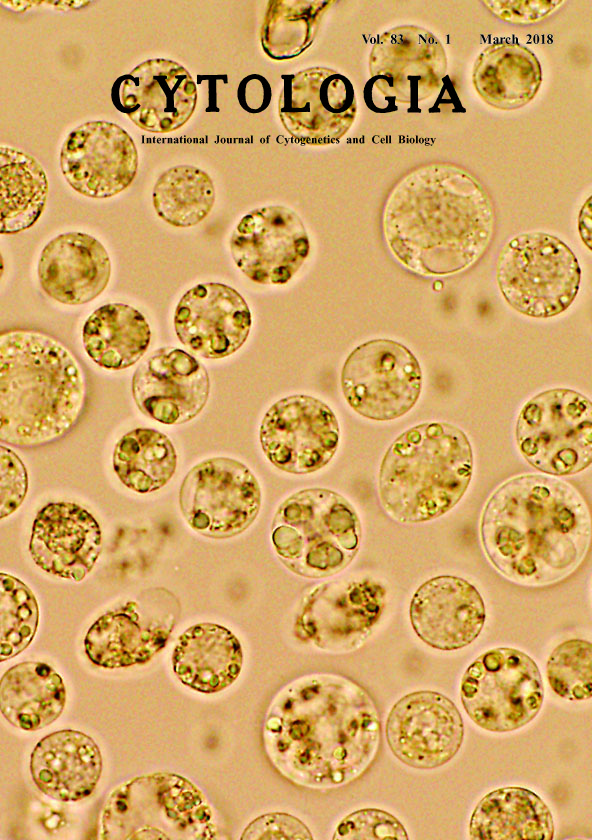 |
|
|---|---|---|
| Vol. 83 No.1 March 2018 | ||
| Technical Note | ||
|
|
||
Isolation of Protoplasts from Suspension Culture of Ceratopteris richardii Seiichiro Hasezawa and Kae Akita* Department of Integrated Biosciences, Graduate School of Frontier Sciences, The University of Tokyo, Kashiwanoha, Kashiwa, Chiba 277–8562, Japan Received November 30, 2017; accepted December 18, 2017 Land plants include morphologically diversified groups. Ferns are thought to offer key advantages for the study of developmental processes leading to complex organs. Ceratopteris richardii is a fern species belonging to the genus Ceratopteris, one of only two genera of the Ceratopteridoideae subfamily of the Pteridaceae (Christenhusz et al. 2011). As C. richardii has been well investigated building a body of knowledge and has been developed as models to be able to analyze their gene functions in development with gene manipulation techniques (Rutherford et al. 2004), we have established the protoplast system from C. richardii cultured cells for an useful experimental tool of plant growth, development, and survival, including cell division, expansive cell growth, biomechanical properties, and stress responses. We obtained a callus derived from spores of C. richardii, further cultured as suspension culture, and succeeded in successive culturing with a rotary shaker. We then developed a procedure for isolating the protoplasts from the cell line, and the protoplast showed the dividing activity. The protoplast system using suspension culture of fern plants is rare, and is thought to be an important tool for the research in cell physiology and genetic engineering of fern plants. Cover figure showed C. richardii protoplasts immediately isolated. The suspension cultures at three days after the transfer were used as the source of protoplasts. The cells were treated with the enzyme solution consisting of 1% Cellulase “ONOZUKA” RS (Yakult Pharm. Ind. Co., Ltd., Tokyo, Japan) and 0.1% pectolyase Y23 (Kyowa Chem. Ind. Co., Ltd., Kagawa, Japan) in 0.5 M mannitol (pH 5.5) at 30°C. The freshly isolated protoplasts were washed with the mannitol solution accompanied by the repeated sequential filtration with stainless filter 500–75 μm pore sizes (Tokyo Screen Co., Ltd., Tokyo, Japan). The protoplasts shows the spherical shapes as a result of completely removing the cell wall. To acquire this image, we used a fluorescence microscope FSX100 (Olympus, Tokyo, Japan) equipped with ×40 objective lens.
Christenhusz, M. J. M., Zhang, X.-C. and Schneider, H. 2011. A linear sequence of extant families and genera of lycophytes and ferns. Phytotaxa 19: 7–54. Rutherford, G., Tanurdzic, M., Hasebe, M. and Banks, J. A. 2004. A systemic gene silencing method suitable for high throughput, reverse genetic analyses of gene function in fern gametophytes. BMC Plant Biol. 4: 6. *Corresponding author, e-mail: kae.akita@gmail.com DOI: 10.1508/cytologia.83.1 |
||