| ON THE COVER | 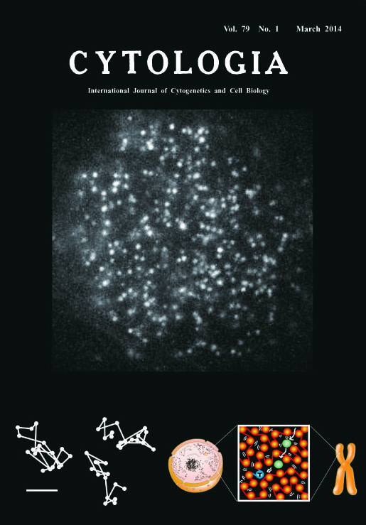 |
|
|---|---|---|
| Vol. 79 No.1 March 2014 | ||
| Technical note | ||
|
|
||
| Single Nucleosome Imaging in Living Human Cells Approximately 2 m of human genomic DNA is organized in the cell. DNA is wrapped around core histones and forms a nucleosome fiber known as “beads-on-a-string”. The nucleosome fiber has long been assumed to be folded into a regular 30-nm chromatin fiber and larger fibers folded into helical structures. However, several lines of evidence, including our recent studies using cryoelectron microscopy and synchrotron X-ray scattering analyses1-4) demonstrated no regular structure >11 nm in interphase chromatin and mitotic chromosomes. These findings suggest that interphase chromatin and mitotic chromosomes consist of compact and irregularly folded nucleosome fibers without a 30-nm chromatin structure (i.e., a polymer melt-like structure). This structure implies a less physically constrained and more locally dynamic state. Nucleosome fibers may thus be constantly moving and rearranging at the local level, which may be crucial for protein factors in the search for genomic DNA. How can we observe such local behavior of the nucleosome in living human cells? To observe single nucleosomes and analyze their local dynamics in living human cells, histone H2B was fused with photoactivatable (PA)-green fluorescent protein (GFP) and expressed in HeLa cells at a very low level.5,6) For single nucleosome imaging, an oblique illumination microscope was used to illuminate a limited thin area within the cell (Nikon laser TIRF microscope system Ti with sapphire 488-nm laser). Generally, PA-GFP exhibits green fluorescence only after activation by a 405-nm laser. However, we unexpectedly found that a small fraction of H2B-PA-GFP in HeLa cells was spontaneously activated without laser stimulation.5,6) The cover image shows a typical single nucleosome image of a living HeLa cell. Each bright dot in the nucleus represents a single H2B-PA-GFP in a single nucleosome. Nucleosome signals in interphase chromatin and mitotic chromosomes in living HeLa cells were recorded at a video rate of ca. 30 ms/frame. Local nucleosome fluctuation was observed (ca. 50 nm movement/30 ms), presumably caused by Brownian motion.5,6) Three representative trajectories of fluorescently tagged single nucleosomes are shown (bottom left; scale bar=100 nm). Combined with a computer simulation, our results suggest that local nucleosome dynamics facilitate chromatin accessibility and play a critical role in scanning genome information.5,6) A scheme of our results is shown (bottom right). In the cell, nucleosome fibers (orange spheres and lines) are irregularly folded and fluctuate. This nucleosome fluctuation assists a protein factor (green sphere) to reach a target sequence in the genome (blue sphere). (Tadasu Nozaki1,2, Ryosuke Imai1,3, Sachiko Tamura1, and Kazuhiro Maeshima1,3*, 1 Structural Biology Center, National Institute of Genetics, Mishima, Shizuoka 411-8540, Japan. 2 Institute for Advanced Biosciences, Keio University, Fujisawa, Kanagawa. 3 Department of Genetics, Sokendai, Mishima, Shizuoka.)
2) Maeshima et al. (2010) Curr. Opin. Cell Biol. 22: 291–297. 3) Nishino et al. (2012) EMBO J. 31: 1644–1653. 4) Joti et al. (2012) Nucleus 3: 404–410. 5) Hihara et al. (2012) Cell Reports 2: 1645–1656. 6) Nozaki et al. (2013) Nucleus 4: 349–356. |
||