| ON THE COVER | 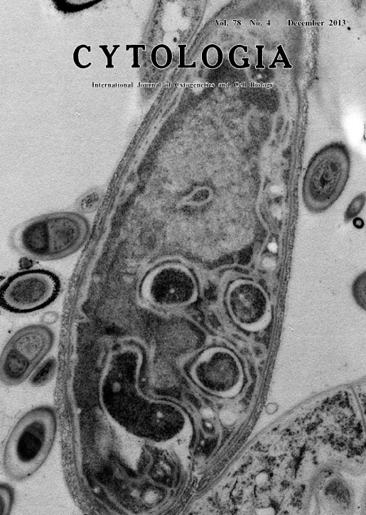 |
|---|---|
| Vol. 78 No.4 December 2013 | |
| Technical note | |
|
|
|
| Ultrathin Sectioning of the Myojin Parakaryote Discovered in the Deep Sea There are only two kinds of organisms on earth: prokaryotes and eukaryotes. Although eukaryotes are considered to have evolved from prokaryotes, there were no previously known intermediate forms between them. The differences in their cellular structures are so vast that the problem of how eukaryotes could have evolved from prokaryotes is one of the greatest enigmas in biology. In 2012, we discovered a unique organism with cellular structures appearing to have intermediate features between prokaryotes and eukaryotes in the deep sea off the coast of Japan (Yamaguchi et al. 2012, Journal of Electron Microscopy 61: 423-431). The organism was 10 μm long and 3 μm in diameter, having more than 100 times volume of Escherichia coli, a common bacterium. It had a large ‘nucleoid,’ consisting of naked DNA fibers, with a single-layered ‘nucleoid’ membrane, and endosymbionts that resemble bacteria, but no mitochondria. Because this organism appears to be a life form distinct from both prokaryotes and eukaryotes but similar to eukaryotes, we named this unique microorganism the ‘Myojin parakaryote’ with the scientific name of Parakaryon myojinensis (“next to (eu)karyote from Myojin”) after the discovery location and its intermediate morphology. The existence of this organism is an indication of a potential evolutionary path between prokaryotes and eukaryotes, and supports the endosymbiotic theory for the origin of mitochondria and the karyogenetic hypothesis for the origin of the nucleus. The Myojin parakaryote was found attached to the chaetae of a scale worm (Polynoidae). The scale worms were collected from the sea bottom at a depth of 1,240 m, and fixed with 2.5% glutaraldehyde in seawater while aboard the ship. The samples were kept on ice and brought to the laboratories of Chiba University. The glutaraldehyde-fixed chaetae and associated microorganisms were cut from the scale worms, sandwiched between two copper discs, and snap-frozen by plunging into melting propane kept in liquid nitrogen. The specimens were freeze-substituted in acetone containing 2% osmium tetroxide at -80°C for 2 days and embedded in epoxy resin (Yamaguchi et al. 2011, Journal of Electron Microscopy 60: 283-287). Serial ultrathin sections were cut, picked up on slit grids, stained with uranyl acetate and lead citrate, and observed with a JEM- 1400 electron microscope (Yamaguchi et al. 2009, Journal of Electron Microscopy 58: 119-128). (Masashi Yamaguchi, Medical Mycology Research Center, Chiba University, 1-8-1 Inohana, Chuo-ku, Chiba 260-8673, Japan) 333-334 |
|