| ON THE COVER | 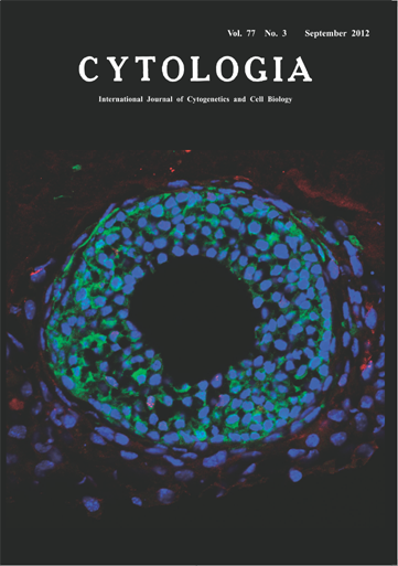 |
|---|---|
| Vol. 77 No.3 September 2012 | |
| Technical note | |
|
|
|
| Theca Cell Layer Formation in Mouse Ovarian Follicle Culture in vitro In mammalian ovaries, oocytes are intimately associated with somatic cells (granulosa cells and theca cells), forming follicles. The theca cells form outermost cell layer of a follicle, surrounding the oocyte and granulosa cells. Although it has been predicted that ovarian interstitial cells (present outside the follicles) are recruited to follicles to form theca cell layer, there has been no experimental evidence to support this prediction. Thus, the mechanism of theca cell layer formation has remained obscure. To clarify the origin of theca cells, we applied a recently developed follicle culture system (Itami et al. 2011, Reproductive Biology and Endocrinology 9: 159–172). In this culture system, follicles are co-cultured with interstitial cells in collagen gel. The follicles, while maintaining their three-dimensional structures, form theca cell layer around them. In order to distinguish between interstitial cells and follicle cells, we used follicles isolated from green fluorescent protein transgenic mouse and interstitial cells prepared from non-transgenic ICR mouse. To analyze cell proliferation, replicating nuclei were labeled with 5-bromo-2′-deoxyuridine (BrdU) for 2 h prior to fixation. The follicles were fixed, embedded in OCT compound, cryo-sectioned (10 μm thick), and the GFP and BrdU were respectively detected by indirect immuno-fluorescent staining. After the cell nuclei were stained with 4, 6-diamidino-2-phenylindole (DAPI), the specimens were examined using a confocal laser scanning microscope system. The picture on the title page represents merged image of GFP (green), BrdU (red), and DAPI (blue), which presents the first experimental evidence for the involvement of interstitial cells in theca cell layer formation. While granulosa cells are all GFP-positive, the theca cell layer (characterized by their flattened cell shape) included a large number of GFP-negative cells as well as a small number of GFP-positive cells, indicating that majority of theca cells comes from interstitial cells while some are derived from the theca cells originally associated with the follicle. Presence of BrdU-positive cells in theca cells suggest that not only cell recruitment but also proliferation of theca cells contribute to the theca cell layer formation. (Saori Itami 1 , Keiko Yasuda 2 , Satoshi Tamotsu 1 and Atsushi Sakai 2 , 1 School of Natural Science and Ecological Awareness, Graduate School of Humanities and Sciences, Nara Women’s University, Kitauoynishi-machi Nara 630–8506, Japan. 2 Department of Biological Sciences 2 , Faculty of Science, Nara Women’s University, Kitauoyanishi-machi, Nara 630–8506, Japan) |
|