| ON THE COVER | 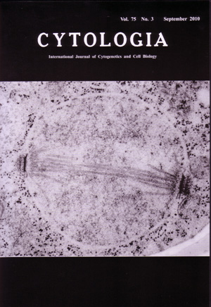 |
|---|---|
| Vol. 75 No.3 September 2010 | |
| Technical note | |
|
|
|
| Meiosis I in Saccharomyces cerevisiae by Rapid-Freeze Electron Microscopy Rapid-freeze electron microscopy of the thin section through a meiosis I nucleus shows the typical spindle structure, which is similar to that seen during mitosis in Saccharomyces cerevisiae. The spindle pole bodies (SPBs) of the budding yeast are complex cylindrical multilayered organelles embedded in the nuclear envelope. The cytoplasmic face of the SPB is the outer plaque, and next to the intermediate line, central plaque with half-bridge and inner plaque. Meiotic prophase is of particular cytological interest because the paired chromosomes are joined along their length by synaptonemal complex, and the duplicated SPBs which are joined by the bridge, remain in side-by-side configuration. The SPBs remain paired throughout meiotic prophase, and then undergo cutting the bridge to make the half-bridge and separation to form the spindle for meiosis I. This separation requires microtubules and results in the generation of a short intranuclear spindle connecting the 2 SPBs. This spindle is composed of kinetochore microtubules and nonkinetochore microtubules which are including continuous pole-to- pole and discontinuous microtubules. The conclusion of meiosis I is most notable for its failure to produce 2 separate nuclei, instead the original nucleus remain single. Then the 2 SPBs each undergo another duplication to produce 2 pairs of duplicated SPBs. Separation of these pairs of SPBs then leads to formation of the 2 spindles for meiosis II within a single nucleus, and then generates 4 ascospores. (Aiko Hirata, Bioimaging Center, Graduate School of Frontier Sciences, The University of Tokyo, Bldg. FSB-601, 5-1-5 Kashiwanoha, Kashiwashi, Chiba 277-8562, Japan) |
|