| ON THE COVER |  |
|---|---|
| Vol. 74 No.4 December 2009 | |
| Technical note | |
|
|
|
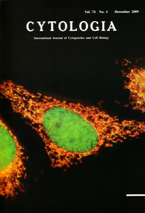
| Visualization of mitochondrial nucleoids in living human cells using SYBR Green I
Mitochondria possess their own genome, the mitochondrial DNA (mtDNA). Human mtDNA is a small, circular DNA molecule, 16.5 kb in length. It encodes 13 polypeptide subunits of respiratory complexes, 22 tRNAs and 2 rRNAs. In mitochondrion, mtDNA molecules associate with various proteins to form compact DNA-protein complexes known as mitochondrial nucleoids (mt-nucleoids). In humans, it is difficult to visualize mt-nucleoids by conventional fluorescent DNA stains such as 4', 6'-diamidino-2- phenylindole (DAPI) and Hoechst because of the small amount of mtDNA per mt- nucleoid. SYBR Green I (Molecular Probes) is a highly sensitive fluorochrome and suitable for the visualization of tiny mt-nucleoids (see Maeda-Sano et al., in this issue). In living HeLa cells, DNA and mitochondria were stained simultaneously with SYBR Green I and MitoTracker Red (Molecular Probes), respectively. The cells grown on the cover slip were incubated in culture medium containing SYBR Green I (diluted 1 : 100,000) and 100 nM MitoTracker Red for 5 min at 37°C in humidified 5 % CO2. The cells were observed under blue excitation with an epifluorescence microscope (BHS-RFC; Olympus). Green fluorescence from SYBR Green I and red fluorescence from MitoTracker Red were simultaneously observed by using a band-path filter. A number of yellow (green plus red) punctate mt-nucleoids in red mitochondria and a green nucleus were observed in each cell (cover image). Most of each mitochondrion contained several mt-nucleoids, whose size was 0.2-0.5 µm. Scale bar, 10 µm. (Sumiko Ozawa1 and Narie Sasaki 1,21 Department of Biology, Faculty of Science, Ochanomizu Unlversity 2-1-1 Ohtsuka, Bunkyo-ku, Tokyo 112-8610, Japan; 2 Division of Biological Science, Graduate School of Sciencc, Nagoya University, Furo-cho, C'hikusa-ku, Nagoya 464-8602, Japan.) |
|