| ON THE COVER | 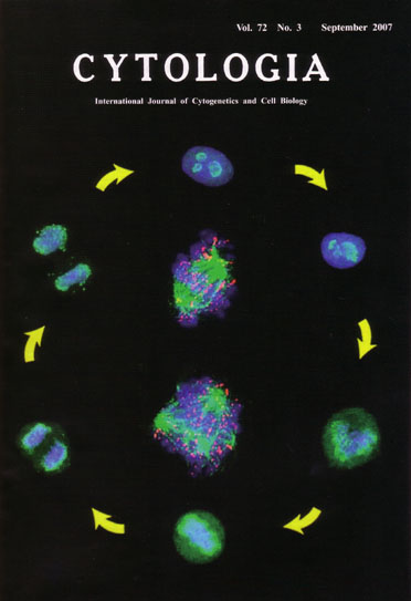 |
|---|---|
| Vol. 72 No.3 September 2007 | |
| Technical note | |
|
|
|
| The chromosome peripheral protein nucleolin locates in the chromosome periphery
and functions in chromosome congression A layer around chromosomes during mitosis was first observed by light microscopy about 80 years ago, and subsequent electron microscopic studies revealed a layer of closely packed fibrils and dense granules around each chromosome. This layer, designated the 'chromosom periphery', appears at prometaphase and disappears at telophase during mitosis. Nucleolin is one of the proteins identified in our proteome analysis of purified human metaphase chromosomes as a chromosome peripheral protein. Nucleolin is known as a multifunctional protein, however, no previous study has demonstrated its functions during mitosis. Recently, Ma N. et al. (Journal of Cell Science, 120, 2091-2105, 2007) demonstrate that the localization pattern of nucleolin throughout the cell cycle. Six cells in clockwise of the cover showed HeLa cells immunostained with antibodies against nucleolin in green and stained with DAPI in blue. At interphase (top), nucleolin is prominently localized at the outer layer of the nucleoli, and present at a lower concentration throughout the nucleoplasm. At prophase (upper right), most of the nucleolin remains in the nucleolar remnants. When the nuclear membrane and nucleoli are disrupted at prometaphase (lower right), nucleolin begins to disperse throughout the cytoplasm and localize at the chromosome periphery. At metaphase (bottom), nucleolin is localized at the chromosome periphery and slightly distribute in the cytoplasm. At anaphase (lower left), most of the nucleolin remains associated with the chromosome periphery, while some becomes packagcd into cytoplasmic particles known as nucleolus-derived foci (NDFs). At telophase (upper right), nucleolin is still associated with chromosomes, and most of it accumulates into NDFs and pre-nucleolar bodies Nucleolin depletion by RNAi mainly induced two types of pronounced defects in chromosome congression: misalignment (upper in the center of the cover) and non-alignment (lower). Two images are nucleolin-depleted cells immunostained with an antibody against α-tubulin in green. a kinetochore marker CREST in red and stained with DAPI in blue. We show that this defect is due to improper kinetochore attachments, resulting in reduced tension, and syntelic attachments of microtubules at the kinetochore. (Ma Nam, Sachihiro Matsunaga, Hideaki Takata, Uchiyama Susumu and Kiichi Fukui, Department of Biotechnology, Graduate School of Engineering, Osaka University, 2-1 Yamadaoka, Suita 565-0871, Osaka, Japan) |
|