| ON THE COVER | 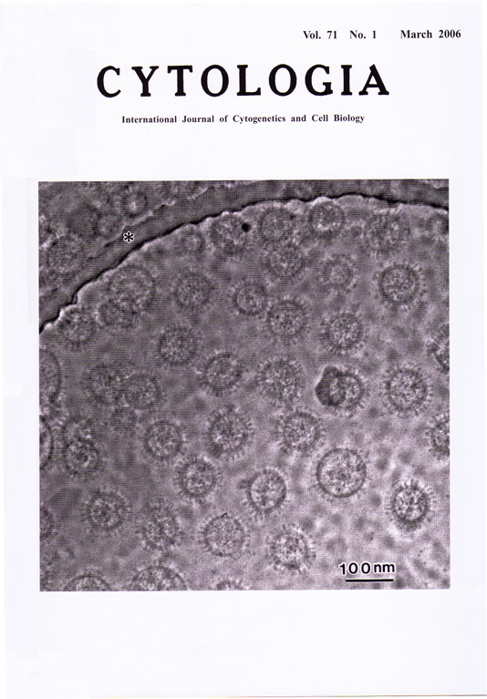 |
|---|---|
| Vol. 71 No.1 March 2006 | |
| Technical note | |
|
|
|
| Phase contrast transmission electron micrograph of rapidly frozen ice-embedded
influenza A virus A suspension of purified influenza A virus in phosphate buffered saline, inactivated with 0.5% formalin, was placed on a perforated carbon grid (*) that was made hydrophilic by glow discharge. Most of the liquid on the grid was removed by blotting with a filter paper, and the grid was rapidly frozen by immersing into liquid ethane kept in liquid nitrogen. With this procedure, very thin layer of frozen virus suspension in a vitrous state was formed. The specimen without staining was transferred in a JEOL JEM3100FFC electron microscope equipped with cryo-stage and Zernike phase contrast phase plate [K. Nagayama, "Phase Contrast Enhancement with Phase Plate in Electron Microscopy", Adv. Imaging Electr. Microsc. 138 (2005) 69-146]. Micrographs were taken at direct magnification of 40,000 at 300kV accelerating voltage using minimum dose system at -271°C with CCD camera. The virions were spherical, 100-120 nm in diameter, had spikes of about 26.3 nm in length, and had lipid bilayer of about 6.4 nm thick. The entities of the spikes are thought to be haemagglutinin and neuraminidase molecules. Note clear virus images with high contrast by phase-contrast effect. (Keishin Sugawara1, Radostin Danev2, Masashi Yamaguchi3, Kiyoto Nishiyama1 and Kuniaki Nagayama2, 1The Chemo-Sero-Therapeutic Research Institute, Kikuchi Research Center, Kyokushi, Kikuchi, Kumamoto 869-1298, Japan, 2Okazaki Institute for Integrative Bioscience, National Institute of Natural Sciences, Myodaiji-cho, Okazaki 444-8787, Japan, 3Research Center for Pathogenic Fungi and Microbial Toxicoses, Chiba University, 1-8-1 Inohana, Chuo-ku, Chiba 260-8673, Japan) |
|
| |
|