| ON THE COVER | 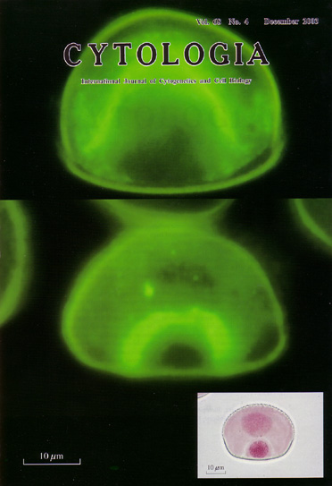 |
|---|---|
| Vol. 68 No.4 December 2003 | |
| Technical note | |
|
|
|
| The mitotic apparatus of Tradescantia paludosa microspores has a peculiar developmental manner related to unequal division
of large vegetative and small generative cells. Microtubules within the
mitotic apparatus were detected under the fluorescence microscope (Nikon
Microphoto-FX) using the anti-α-tubulin immunofluorescence methods. The
microspores were fixed with 3.7% paraformaldehyde in a potassium phosphate
buffer solution, pH 6.8. The mouse monoclonal anti-α-tubulin antibody (Amersham)
and goat anti-mouse IgG-FITC (Tago) were used, respectively, as the first
and second antibodies. Throughout the microspore division, mitotic apparatuses were organized asynchronously and/or asymmetrically. From prophase to prometaphase, the spindle began to construct itself in the vegetative pole preceding the generative pole. The prophase spindles on the vegetative pole side were in the form of domes or cones that lacked the top (upper panel). The half-spindles were asymmetric at the metaphase and the anaphase. The phragmoplast developed curving toward the generative pole during telophase (middle panel). It first grew toward the long axis of the ellipsoidal-shaped microspore: and after it arrived at the inner membrane of the microspore, it again curved past the generative nucleus toward the short axis. A new cell plate was formed in the areas through which phragmoplast travelled. A large vegetative cell and a smal1 hemispherical generative cell were also formed (lower panel; aceto-carmine staining) (see Terasaka, O., Niitsu, T. 1990: Bot. Mag. Tokyo 103: 133-142, Terasaka, O., Niitsu, T. 1995: Protoplasma 189: l87-193). (Osamu Terasaka, Biological Laboratory, Department of Natural Sciences, Jikei University School of Medicine, Kokuryo, C'hofu, Tokyo, 182-8570, Japan) |
|