| ON THE COVER | 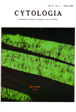 |
|---|---|
| Vol. 67 No.1 March 2002 | |
| Technical note | |
|
|
|
| Cortical microtubules in living epidermal cells
of Arabidopsis. We have obtained a transgenic Arabidopsis plant that stably expresses a GFP-α-tubulin-6 fusion protein (Ueda et
al. 1999). With this plant, the GFP fluorescence of microtubules can be detected by a laser fluorescence microscope, and the shape and distribution
pattern of microtubules in the living cells can be visualized. The establishment of the transgenic Arabidopsis plant is as follows. An Arabidopsis α-tubulin TUA6 cDNA in pZL1 was subcloned into pBluescript KS-between SalI and NotI sites, and an NaeI site was introduced in front of the translation initiation Met by PCR amplification. The stop codon of a soluble-modified red-shifted version of GFP (smRS-GFP) was replaced with an NaeI site by PCR amplification. The cDNAs of TUA6 and smRS-GFP were then cut with Nael and Sacl, and with BamHI and Nael, respectively, and ligated together between BamHI and Sac1 sites in pBluescript KS- .The resulting plasmid contained an in-frame fusion between smRS-GFP and TUA6, connected by an Ala-Gly linker encoded in the Nael site. The JFP-TUA6 fusion sequence was then inserted between the BamHI and Sacl sites in pBI 121 so that the fusion protein is expressed under the 35S prcunoter.A thaliana ecotypc Columbia plants with a gl1 marker were vacuum infiltrated with the Agrobacterium tumefaciens pGV2260 strain containing the above construct. Tl seedlings were screencd for kanamycin resistance (30mg/L). Transgenic lines homozygous for kanamycin resistance were selected at T2 and T3 generations, and used for analysis. Ref., Ueda, K. and Matsuyama. T. (2000) Rearrangement of cortical microtubules from transverse to oblique or longitudinal in living cells of transgenic Arabidopsis thaliana. Protoplasma 213: 28-38. (Katsumi Ueda: Department of Biological Sciences. Nara Women's University, Nara 630-8506, Japan) |
|