[-> contents]
[ Document Identification Number : DIN00032503 ]
Proceedings of the 2nd Symposium of the 'Color' of Digital Imaging in Biology and Medicine,
3.1-3.4, 2000.03.25
<http://biocolor.umin.ac.jp/sympo200004/proc13.pdf>
第2回デジタル生体医用画像の「色」シンポジウム

パネルディスカッション「デジタル生体医用画像の色のあり方」
―その1:要素技術からのアプローチ―

肌の絶対分光反射率画像の計測とその成分分析への応用

津村 徳道*1 (tsumura@ics.tj.chiba-u.ac.jp)
Francisco H. Imai*2、三宅 洋一*1
*1千葉大学工学部情報画像工学科、*2ロチェスター工科大MCSL


The 2nd Symposium of the 'Color' of Digital Imaging in Biology and Medicine

Panel Discussion "What the 'Color' of Digital Imaging in Biology and Medicine Should Be"
- Part 1 : Approaches From the Aspects of Key Technologies -

Measurement of Absolute Spectral-reflectance-image of Skin and
the Application of Component Analysis


Norimichi TSUMURA*1 (tsumura@ics.tj.chiba-u.ac.jp)
Francisco H. Imai*2, Yoichi MIYAKE*1
*1Deptartment of information and Image Sciences, Chiba University
*2MCSL, Rochester Institute of Technology

Summary
Mapping the oxygen saturation of blood and melanin in skin is expected to give useful information for skin diagnosis. We have already proposed a technique to estimate the map of skin components from spectral reflectance image obtained by multi-band images based on the optical scattering analysis. The spectral reflectance image is a result of dividing the spectral image of object by that of absolute white reference plane. However, the shape of skin is not a two-dimensional plane, so that the required absolute spectral reflectance is not obtained in the wide region of skin by the conventional method.
In this research, the absolute spectral reflectance and the normal vector of the surface were obtained by using the photometric stereo technique. In the photometric stereo technique, more than three illuminants were used, and an image was taken by each illuminant. The pixel values in several obtained images were used to calculate the absolute spectral reflectance and the normal vector of the surface on the corresponding pixel. The index finger where the second joint was bound by a string was captured and analyzed by the proposed technique. The results show that the components are extracted for the wide range of the finger.

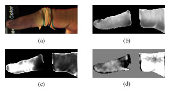
 皮膚の酸素飽和率分布やメラニン色素の分布が得られれば、それらは肌疾患の診断や治療に有効であると考えられる。これまで我々は、肌のマルチバンド撮影により得られた分光反射率画像を用いて、光散乱解析に基づき肌の酸素飽和率分布やメラニン色素分布を推定する手法を提案してきた1。 ここで、分光反射率画像は、肌が2次元平面物体であると仮定することにより、撮影された肌の分光画像から肌面と法線方向を同一とする参照白色板の分光画像を除算して得られる。しかし、実際には肌は3次元物体であるため、色素分布の推定に必要である絶対的な分光反射率が得られる領域は限られている。参照板と肌面の法線方向が異なる領域では、絶対的な分光反射率に法線方向のずれに応じて定数倍された相対的な分光反射率しか得られない。
 一方、近年の計算機処理能力の急速な発展により、物体の3次元形状や色、質感などの情報を、実際に撮影された画像などからコンピュータビジョン技術により抽出し、コンピュータグラフィック技術により任意の視点、照明環境下で表示するImage Based Renderingが現在盛んに研究されている。
 そこで本研究では、コンピュータビジョン技術の一つであるPhotometric Stereo法を用いて、肌の各点の絶対分光反射率と面の法線方向を抽出し、また抽出結果や成分分析の結果をコンピュータグラフィック技術を用いて任意の視点、照明環境下で表示した。今回用いたPhotometric Stereo法では、物体の反射特性として完全拡散面(Lambertian面)を仮定することにより、3方向以上の位置から別々に照明された複数枚の画像の画素値から、各点の絶対分光反射率と面の法線方向を計測することが出来る。
 実際に、第2関節においてひもを用いて止血した人差し指に対する実験を行った。今回、4方向の照明位置から順に撮影した。各照明における照度ムラは参照白色板を用いて近似的に校正した。図1に、得られた成分分析の結果を示す。絶対分光反射率から肌の広範囲にわたる酸素飽和率分布やメラニン色素分布などを推定することができている。また、図2に、得られた各点の法線方向から3次元形状を復元し、コンピュータグラフィック技術により撮影時と異なる視点で表示した結果を示す。 指の3次元形状と成分分析の結果を合わせて見ることができる。

図1 第2関節においてひもを用いて止血した人差し指の成分分析結果
(a)推定した絶対分光反射率をもとにD65光源化で一様照明された場合を仮定して再現した画像、(b)メラニン分布、(c)全ヘモグロビン分布、(d)酸素飽和率分布

Figure 1  The result of component analysis for index finger where the second joint was bound by string.
(a) original image, (b) melanin component, (c) total hemoglobin, (d) oxygen saturation


図2 コンピュータグラフィックス技術を用いた酸素飽和率分布の
撮影時と異なる視点からの表示
Figure 2  Display of oxygen saturation by computer graphics technique.


 今後、肌の表面反射光などが含まれたより複雑な反射特性を考慮し、また計測結果に大きく影響を及ぼす照明ムラの校正方法を改善する必要がある。しかし、今回の研究により、コンピュータビジョン技術を医用画像計測に用いる有効性を示すとともに、コンピュータグラフィック技術と組み合わせて用いることにより遠隔医療などにも適用可能な新しい医用画像システムの可能性が示された。

謝辞 貴重なご意見を頂いたロチェスター大学のM. A. Kriss教授, K. A. Parker教授, K. Kutulacos教授に感謝いたします。

参考文献
【1】川渕、津村、羽石、三宅:第46回応用物理学関係連合講演会講演集、1056、1999年