[-> contents]
[ Document Identification Number : DIN01022815 ]
Digital Color Imaging in Biomedicine (in press), 2001.02.28 (draft)
<http://biocolor.umin.ac.jp/book200102/din01022815.pdf>
Digital Color Imaging in Biomedicine (in press), 2001.02.28 (draft)
<http://biocolor.umin.ac.jp/book200102/din01022815.pdf>
|
Digital Imaging in Forensic Medicine
Kiyoshi MATSUI*1 (matsuik@art.osaka-med.ac.jp) *1Department of Forensic Medicine, Osaka Medical College |
|
Abstract: Superimposing an image of a skull on an image of a face is one of the most effective methods for identifying a deceased individual from his/her skeleton. Both ordinary methods using photography and video require an actual skull, but the body of the deceased must be cremated within a certain period.
In this paper, a new method is introduced, in which pictures of possible persons' faces and a skull to be inspected are digitized and superimposed using advanced image processing software running on a personal computer. This software has adequate functions to inspect essential characteristics required to decide whether or not the face agrees with the skull. This method is very useful and easy to use, but attention should be paid to avoid artificial or unintentional manipulation of digitized images. |
|
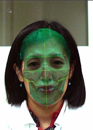
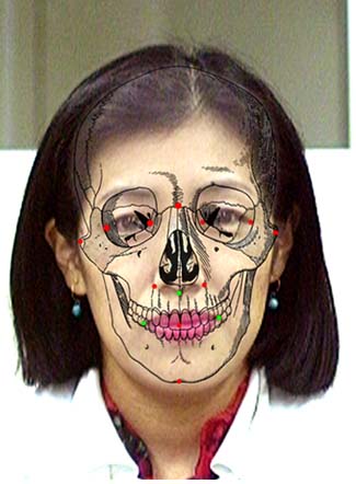
1. Introduction Among autopsies carried out in department of forensic medicine, judicial autopsy has the forensic aspect of collecting evidence in addition to the medical aspect of elucidating the cause of death, because findings obtained during autopsy may be of legal value later. Those findings are collected in two forms now. One is literally recorded information on properties such as shape, color, or dimension that are usually dictated by a doctor and the other form is photographic record which conserves the properties of the cadaver as they are. A subjective point of view is inevitably included into the former form whereas photographs can record visual perception as objectively as possible. Photographs are thus very important in forensics because in some court cases only photographs are available for judgment instead of the original. Color is one of the most important findings because it reflects critically the cause of death in certain cases such as carbon monoxide intoxication or suggests aging of subcutaneous hemorrhage. Therefore, illumination in the autopsy room should be adjusted to reflect the natural color of the objects. Digitized images are increasingly used in medical fields and strict device calibration is prerequisite for utilizing digitized images instead of photographs for storage of autopsy findings. Human skeletal remains are also subjected to expert evaluation. In many of such cases the person's identification remains unknown as well as the cause of death. Therefore, we should collect as many kinds of information as possible from such skeletal remains: physical properties, sex, age, bone injuries, characteristics of teeth, postmortem period, blood types, or DNA types. Among them critical information for identifying the remains is mainly provided by cranio-facial properties which are used for superimposition. We present here digital superimposition of skulls and photographic portraits performed on a Macintosh PowerPC computer using Adobe Photoshop software. The use of antemortem photographic records of facial features to identify human skeletal remains has been well established over the past 50 years. Comparison of the correctly enlarged photograph with the photographed skull allow us to evaluate the anatomical consistency between them. The identification process has become sophisticated, from photographs in the beginning to video-mediated methods later, but the latter method requires expensive and complex equipment. Both methods need a real skull during the process. Many skulls are subjected to cremation after a certain period because of custody difficulties before the photographs of the relevant person are obtained. Therefore, photographic records during autopsy only can remain to be used for superimposition. Superimposition performed using computer graphics thus plays a major role in personal identification. 2. Materials and methods Recent developments in computer technology have enabled us to handle any kinds of images such as illustrations, photographs, or animations as digital data on personal computers using high-performance image-processing technology that was formerly accessible exclusively to professionals. We performed the superimpose method by using the overlay function of Adobe Photoshop on a Macintosh personal computer in this study. Some cases where portraits of the relevant person were available they were selected from autopsies of skeletal remains. We used those portraits which were photographed in such a way where the angle and direction looked most like the photographs taken during the autopsy when the candidates of the remains were unknown. Both series of photographs from antemortem portrait and postmortem skull were digitized into the computer using a flatbed scanner. The digital image of the skull was layered over the digital image of the portrait in the background. The same anatomical points described below were marked on both images. The image of the skull was then moved using the "hand tool" of Photoshop to match the points marked on the skull layer with those of the image of the portrait layer. Major anatomical points used as examination criteria for assessment of anatomical consistency between the images of portrait and skull were as follows: Vertex, Nasion, Subnasale, Stomion, Gnathion, Zygion, Entokanthion, Ektokanthion, and Alare. The differences mainly in angle between the skull and the portrait were minimized by changing magnification and rotating the skull image with the transparency of the layer. Anatomical consistency was then examined for the points listed above. Successfully superimposed image layers were merged after deleting unused images and were finally printed out. |
 |
 |
|
3. Results and discussion
High-performance image-processing software enables us to superimpose two series of photographs, antemortem portraits and photographs of a postmortem skull, both of which were taken in different angles and directions. The process begins with pasting the digital image into layers and then the differences such as angle between the two series of photographs were minimized using the "hand tool" of Photoshop. Finally we assessed the consistency of both series of photographs by comparing the marked points the whole contour on the layered images. A higher version of Photoshop with the addition of further functions makes itself more versatile in image-processing including superimpose method. However, it should be noted that the original image might be critically deformed unintentionally and the validity of the superimposition might be jeopardized by over-processing. Comparing the processed image from the portrait used in this study with the image from the other skull (i.e., a different person) showed inconsistencies at many mark points, indicating the high specificity of the method. However, final judgment requires other data such as blood types, dental information, or DNA types in addition to these superimposition techniques. Acknowledgments I am most grateful to Prof. Koichi Suzuki, Department of Forensic Medicine, Osaka Medical College, for his valuable advice. |