[-> contents]
[ Document Identification Number : DIN01022814 ]
Digital Color Imaging in Biomedicine, 77-78, 2001.02.28
<http://biocolor.umin.ac.jp/book200102/din01022814.pdf>
Digital Color Imaging in Biomedicine, 77-78, 2001.02.28
<http://biocolor.umin.ac.jp/book200102/din01022814.pdf>
|
From the Standpoint of Neurosurgery
Hiroshi ISEKI*1 (hiseki@nij.twmu.ac.jp) *1Department of Neurosurgery, Neurological Institute, Tokyo Women's Medical University |
|
In neurosurgery, one of the outstanding applications of advanced medical technology is minimally invasive surgery, which will bring great reduction in both patients' burdens and medical expenditures. The essential elements to realize this are (1) substitution of human hands which can accurately manipulate the object tissue, (2) substitution of human eyes to identify and observe the object of the operation, (3) visual information superimposed on the image of the object which provides navigation to assist the surgeon in the operation. Because endoscopic images give fundamental visual information of the system, they must have high-fidelity color (Fig. 1).
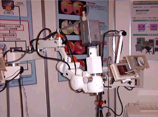
In microsurgery, stereoscopic video microscope systems equipped with a flat panel display and a video camera are replacing ordinary microscopes for microsurgery. With these systems, a surgeon can sit freely in a comfortable position during the operation, all staff members engaged in the operation can share the same images as the operator watches, and all information required for the operator can be visualized on the same display for microscopic images. In addition, an augmented virtual reality technology called HivisCAS can superimpose 3-D maps made for navigation of the operation upon real objects. In these systems, realistic color images are also required (Fig. 2). Solutions for visualizing the image acquired using invisible light, reproducing the feel of a material in a virtual space, and reproducing the same color using different illumination and different monitors, should be pursued to meet the clinical requirements as shown here. |
 |
 |